Structural and dynamic characterization of the hexa-coordinated globin from Spisula solidissima.
Pesce, A., Barmpidi, K., Dewilde, S., Estarellas, C., Moens, L., Bolognesi, M., Luque, F.J., Nardini, M.(2023) J Inorg Biochem 246: 112289-112289
- PubMed: 37354606 Search on PubMed
- DOI: https://doi.org/10.1016/j.jinorgbio.2023.112289
- Primary Citation Related Structures:
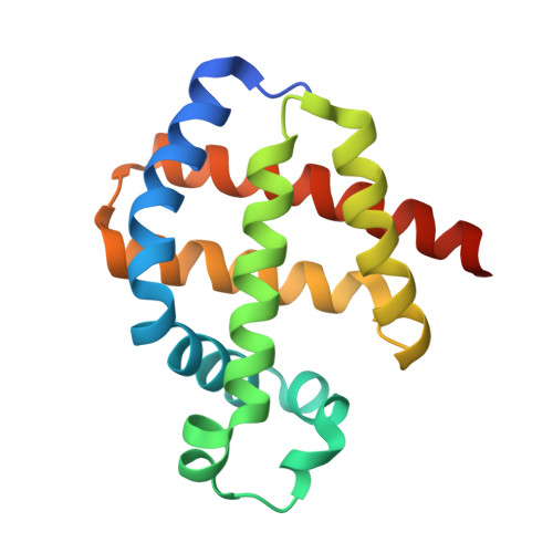
8OUP - PubMed Abstract:
High energy consumption in the nervous system requires a continuous supply of O 2 . This role is assisted by proteins from the globin super-family in the nerve cells of invertebrates, where 'nerve hemoglobins' (nHbs) are mainly present at mM concentrations and exhibit oxygen affinities comparable to those of vertebrate myoglobins. To gain insight into the structural bases of this function, we report the crystal structure of nHb from the Atlantic surf clam Spisula solidissima (SsHb), previously suggested to display a bis-histidyl hexa-coordinated heme in the deoxy state, high O 2 affinity, and ligand binding cooperativity when assayed in situ. The crystallized protein forms a dimer through packing of a 4-helix bundle involving helices E and F of each subunit. The SsHb 'classic' globin fold displays bis-histidyl (His71(E7) and His103(F8)) hexa-coordination of the heme-Fe atom, with structural and dynamics variations found in the inter-helix hinge regions. Molecular Dynamics simulations of both monomeric and dimeric species in the bis-histidyl hexa-coordinated, deoxy penta-coordinated, and O 2 -bound hexa-coordinated states reveal distinct structural rearrangements at the interface between subunits in the dimer; these would affect the magnitude of the conformational fluctuations observed between monomer and dimer, and the topology of cavities within the protein matrix and at the interface. These results point to a distal site opening mechanism allowing access of the exogenous ligand to the heme and cast hypotheses on the dimer interface structural and dynamic properties that may support ligand binding cooperativity in dimeric SsHb.
- Department of Physics, University of Genova, Via Dodecaneso 33, I-16146 Genova, Italy.
Organizational Affiliation: