8ORD

Cryo-EM map of zebrafish cardiac F-actin


Experimental Data Snapshot

  • Method: ELECTRON MICROSCOPY
  • Resolution: 3.90 Å
  • Aggregation State: FILAMENT 
  • Reconstruction Method: SINGLE PARTICLE 

wwPDB Validation   3D Report Full Report


This is version 1.1 of the entry. See complete history


Literature

Zebrafish as a model for cardiac disease; Cryo-EM structure of native cardiac thin filaments from Danio Rerio.

Bradshaw, M.Squire, J.M.Morris, E.Atkinson, G.Richardson, R.Lees, J.Caputo, M.Bigotti, G.M.Paul, D.M.

(2023) J Muscle Res Cell Motil 44: 179-192

  • DOI: https://doi.org/10.1007/s10974-023-09653-5
  • Primary Citation of Related Structures:  
    8ORD

  • PubMed Abstract: 

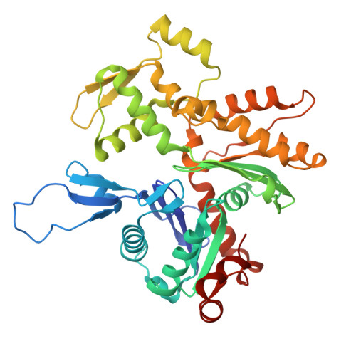
    Actin, tropomyosin and troponin, the proteins that comprise the contractile apparatus of the cardiac thin filament, are highly conserved across species. We have used cryo-EM to study the three-dimensional structure of the zebrafish cardiac thin and actin filaments. With 70% of human genes having an obvious zebrafish orthologue, and conservation of 85% of disease-causing genes, zebrafish are a good animal model for the study of human disease. Our structure of the zebrafish thin filament reveals the molecular interactions between the constituent proteins, showing that the fundamental organisation of the complex is the same as that reported in the human reconstituted thin filament. A reconstruction of zebrafish cardiac F-actin demonstrates no deviations from human cardiac actin over an extended length of 14 actin subunits. Modelling zebrafish homology models into our maps enabled us to compare, in detail, the similarity with human models. The structural similarities of troponin-T in particular, a region known to contain a hypertrophic cardiomyopathy 'hotspot', confirm the suitability of zebrafish to study these disease-causing mutations.


  • Organizational Affiliation

    Physiology, Pharmacology and Neuroscience, University of Bristol, Bristol, UK.


Macromolecules
Find similar proteins by:  (by identity cutoff)  |  3D Structure
Entity ID: 1
MoleculeChains Sequence LengthOrganismDetailsImage
Actin, alpha 1b, skeletal muscle
A, B, C, D, E
377Danio rerioMutation(s): 0 
UniProt
Find proteins for F1RCB6 (Danio rerio)
Explore F1RCB6 
Go to UniProtKB:  F1RCB6
Entity Groups  
Sequence Clusters30% Identity50% Identity70% Identity90% Identity95% Identity100% Identity
UniProt GroupF1RCB6
Sequence Annotations
Expand
  • Reference Sequence
Small Molecules
Ligands 2 Unique
IDChains Name / Formula / InChI Key2D Diagram3D Interactions
ADP
Query on ADP

Download Ideal Coordinates CCD File 
F [auth A],
H [auth B],
J [auth C],
L [auth D],
N [auth E]
ADENOSINE-5'-DIPHOSPHATE
C10 H15 N5 O10 P2
XTWYTFMLZFPYCI-KQYNXXCUSA-N
PO4
Query on PO4

Download Ideal Coordinates CCD File 
G [auth A],
I [auth B],
K [auth C],
M [auth D],
O [auth E]
PHOSPHATE ION
O4 P
NBIIXXVUZAFLBC-UHFFFAOYSA-K
Experimental Data & Validation

Experimental Data

  • Method: ELECTRON MICROSCOPY
  • Resolution: 3.90 Å
  • Aggregation State: FILAMENT 
  • Reconstruction Method: SINGLE PARTICLE 

Structure Validation

View Full Validation Report


Entry History & Funding Information

Deposition Data


Funding OrganizationLocationGrant Number
British Heart FoundationUnited KingdomFS/14/18/30711

Revision History  (Full details and data files)

  • Version 1.0: 2023-08-02
    Type: Initial release
  • Version 1.1: 2023-10-11
    Changes: Data collection, Database references