Structural and thermodynamic insights into antibody light chain tetramer formation through 3D domain swapping.
Sakai, T., Mashima, T., Kobayashi, N., Ogata, H., Duan, L., Fujiki, R., Hengphasatporn, K., Uda, T., Shigeta, Y., Hifumi, E., Hirota, S.(2023) Nat Commun 14: 7807-7807
- PubMed: 38065949 Search on PubMedSearch on PubMed Central
- DOI: https://doi.org/10.1038/s41467-023-43443-4
- Primary Citation Related Structures:
8KAD - PubMed Abstract:
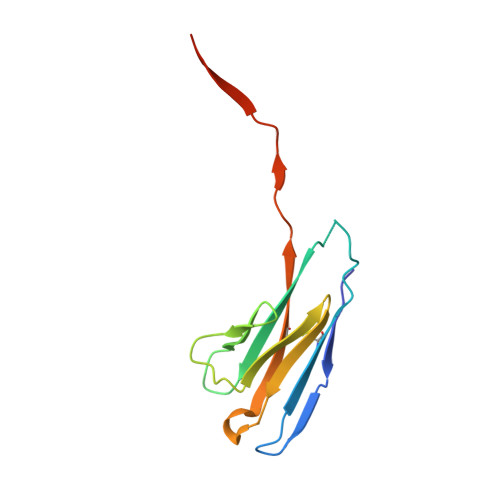
Overexpression of antibody light chains in small plasma cell clones can lead to misfolding and aggregation. On the other hand, the formation of amyloid fibrils from antibody light chains is related to amyloidosis. Although aggregation of antibody light chain is an important issue, atomic-level structural examinations of antibody light chain aggregates are sparse. In this study, we present an antibody light chain that maintains an equilibrium between its monomeric and tetrameric states. According to data from X-ray crystallography, thermodynamic and kinetic measurements, as well as theoretical studies, this antibody light chain engages in 3D domain swapping within its variable region. Here, a pair of domain-swapped dimers creates a tetramer through hydrophobic interactions, facilitating the revelation of the domain-swapped structure. The negative cotton effect linked to the β-sheet structure, observed around 215 nm in the circular dichroism (CD) spectrum of the tetrameric variable region, is more pronounced than that of the monomer. This suggests that the monomer contains less β-sheet structures and exhibits greater flexibility than the tetramer in solution. These findings not only clarify the domain-swapped structure of the antibody light chain but also contribute to controlling antibody quality and advancing the development of future molecular recognition agents and drugs.
- Division of Materials Science, Graduate School of Science and Technology, Nara Institute of Science and Technology, 8916-5 Takayama, Ikoma, Nara, 630-0192, Japan.
Organizational Affiliation: