8JLY | pdb_00008jly

Structure of nanobody in complex with alpha-synuclein peptide


Experimental Data Snapshot

  • Method: X-RAY DIFFRACTION
  • Resolution: 1.29 Å
  • R-Value Free: 
    0.186 (Depositor), 0.186 (DCC) 
  • R-Value Work: 
    0.161 (Depositor), 0.161 (DCC) 
  • R-Value Observed: 
    0.163 (Depositor) 

Starting Model: experimental
View more details

wwPDB Validation 3D Report Full Report

Validation slider image for 8JLY

This is version 1.2 of the entry. See complete history

Literature

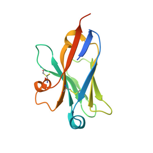
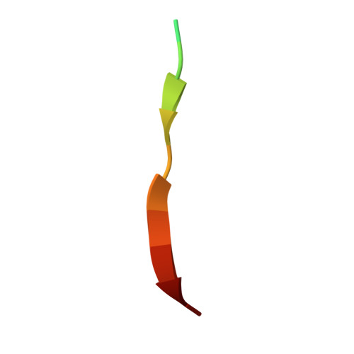
Structural insights into the unique recognition module between alpha-synuclein peptide and nanobody.

Islam, Z.Vaikath, N.N.Hmila, I.El-Agnaf, O.M.A.Kolatkar, P.R.

(2024) Protein Sci 33: e4875-e4875

  • DOI: https://doi.org/10.1002/pro.4875
  • Primary Citation Related Structures: 
    8JJV, 8JLY

  • PubMed Abstract: 

    Nanobodies are single-domain fragments of antibodies with comparable specificity and affinity to antibodies. They are emerging as versatile tools in biology due to their relatively small size. Here, we report the crystal structure of a specific nanobody Nbα-syn01, bound to a 14 amino acid long peptide of α-synuclein (αSyn), a 140-residue protein whose aggregation is associated with Parkinson's disease. The complex structure exhibits a unique binding pattern where the αSyn peptide replaces the N-terminal region of nanobody. Recognition is mediated principally by extended main chain interaction of the αSyn peptide and specificity of the interaction lies in the central 48-52 region of αSyn peptide. Structure-guided truncation of Nbα-syn01 shows tighter binding to αSyn peptide and improved inhibition of α-synuclein aggregation. The structure of the truncated complex was subsequently determined and was indistinguishable to full length complex as the full-length form had no visible electron density for the N-terminal end. These findings reveal the molecular basis for a previously unobserved binding mode for nanobody recognition of α-synuclein, providing an explanation for the enhanced binding, and potential for an alternate framework for structure-based protein engineering of nanobodies to develop better diagnostic and therapeutic tools.


  • Organizational Affiliation
    • Diabetes Center, Qatar Biomedical Research Institute, Hamad Bin Khalifa University, Qatar Foundation, Doha, Qatar.

Macromolecule Content 

  • Total Structure Weight: 15.49 kDa 
  • Atom Count: 1,045 
  • Modeled Residue Count: 120 
  • Deposited Residue Count: 144 
  • Unique protein chains: 2

Macromolecules

Find similar proteins by:|  3D Structure
Entity ID: 1
MoleculeChains  Sequence LengthOrganismDetailsImage
Nanobody130Camelus dromedariusMutation(s): 0 
Find similar proteins by:  Sequence   |   3D Structure  
Entity ID: 2
MoleculeChains  Sequence LengthOrganismDetailsImage
alpha-synuclein peptide14Homo sapiensMutation(s): 0 
UniProt & NIH Common Fund Data Resources
Find proteins for P37840 (Homo sapiens)
Explore P37840 
Go to UniProtKB:  P37840
PHAROS:  P37840
GTEx:  ENSG00000145335 
Entity Groups
Sequence Clusters30% Identity50% Identity70% Identity90% Identity95% Identity100% Identity
UniProt GroupP37840
Sequence Annotations
Expand
Reference Sequence

Small Molecules

Ligands 1 Unique
IDChains Name / Formula / InChI Key2D Diagram3D Interactions
GOL

Query on GOL


Download:Ideal Coordinates CCD File
C [auth A]GLYCEROL
C3 H8 O3
PEDCQBHIVMGVHV-UHFFFAOYSA-N

Experimental Data & Validation

Experimental Data

  • Method: X-RAY DIFFRACTION
  • Resolution: 1.29 Å
  • R-Value Free:  0.186 (Depositor), 0.186 (DCC) 
  • R-Value Work:  0.161 (Depositor), 0.161 (DCC) 
  • R-Value Observed: 0.163 (Depositor) 
Space Group: P 2 21 21
Unit Cell:
Length ( Å )Angle ( ˚ )
a = 30.474α = 90
b = 41.906β = 90
c = 86.352γ = 90
Software Package:
Software NamePurpose
PHENIXrefinement
PHENIXrefinement
XDSdata reduction
XDSdata scaling
PHASERphasing

Structure Validation

View Full Validation Report


Entry History 

& Funding Information

Deposition Data


Funding OrganizationLocationGrant Number
Qatar FoundationQatar--

Revision History  (Full details and data files)

  • Version 1.0: 2024-01-10
    Type: Initial release
  • Version 1.1: 2024-02-14
    Changes: Database references
  • Version 1.2: 2024-10-23
    Changes: Structure summary