Modulating Chromophore Flexibility in GEVIs through Threonine-Based Molecular Switches Reveals an Influence of Membrane Curvature on Protein Activity.
Leong, L.M., Shin, S.C., Frankiv, N., Rhee, J.K., Kim, H., Seong, J., Woo, J., Han, K., Storace, D.A., Baker, B.J.(2025) ACS Sens 10: 8395-8410
- PubMed: 41123309 Search on PubMed
- DOI: https://doi.org/10.1021/acssensors.5c01748
- Primary Citation Related Structures:
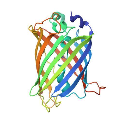
8JKC, 8JKG, 8JKI, 8JL2, 8JL5, 8JL6, 8JL7, 8JLL, 8JLM, 8JLS, 8JLT, 8JLU - PubMed Abstract:
Many genetically encoded voltage indicators (GEVIs) rely on fluorescent protein (FP) domains to report changes in membrane potential. Rapid and reversible disruption of steady-state fluorescence during voltage sensor activation revealed transient conformational changes near the chromophore in the FP domain, implicating chromophore flexibility as a potential mechanism of signal modulation. Substitution of a bulky phenylalanine near the chromophore with threonine (F165T) introduced a distinct secondary component in the fluorescence response, consistent with increased chromophore mobility. This effect was tunable: an external, directionally polarized offset (164 T /166F) reoriented the internal threonine side chain, restoring steric hindrance and eliminating the secondary signal. Thus, threonine can function as a context-sensitive molecular switch shaped by β-can surface chemistry. A second internal threonine (T203) also acted as a molecular switch under modified external conditions, generating a secondary signal that is susceptible to membrane curvature during depolarization suggesting that plasma membrane geometry can modulate GEVI activity under permissive conformational states. Crystal structures of Super Ecliptic pHluorin (SE), SE A227D, and a new FP variant revealed that external residues can influence internal side chain orientation. In the new variant, pH-dependent rearrangement of the seventh β-strand dramatically repositions D147 from the interior interacting with the chromophore to the external surface, while H148 shifts from the exterior to interact with the chromophore in alkaline conditions. These insights led to the development of a new GEVI, Ulla, which inverts the polarity of the optical signal─becoming brighter upon depolarization─displays reduced pH sensitivity in the physiological range, and performs reliably under low-light, high-speed imaging conditions in vitro and in vivo using widefield and 2-photon microscopy. Together, these findings present a new approach to modulating chromophore behavior offering broad potential for FP-based biosensor development.
- Brain Science Institute, Korea Institute of Science and Technology, Seoul 02792, Republic of Korea.
Organizational Affiliation: