Molecular mechanism for endo-type action of glycoside hydrolase family 55 endo-beta-1,3-glucanase on beta 1-3/1-6-glucan.
Ota, T., Saburi, W., Tagami, T., Yu, J., Komba, S., Jewell, L.E., Hsiang, T., Imai, R., Yao, M., Mori, H.(2023) J Biological Chem 299: 105294-105294
- PubMed: 37774972 Search on PubMedSearch on PubMed Central
- DOI: https://doi.org/10.1016/j.jbc.2023.105294
- Primary Citation Related Structures:
8JHH - PubMed Abstract:
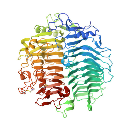
The glycoside hydrolase family 55 (GH55) includes inverting exo-β-1,3-glucosidases and endo-β-1,3-glucanases, acting on laminarin, which is a β1-3/1-6-glucan consisting of a β1-3/1-6-linked main chain and β1-6-linked branches. Despite their different modes of action toward laminarin, endo-β-1,3-glucanases share with exo-β-1,3-glucosidases conserved residues that form the dead-end structure of subsite -1. Here, we investigated the mechanism of endo-type action on laminarin by GH55 endo-β-1,3-glucanase MnLam55A, identified from Microdochium nivale. MnLam55A, like other endo-β-1,3-glucanases, degraded internal β-d-glucosidic linkages of laminarin, producing more reducing sugars than the sum of d-glucose and gentiooligosaccharides detected. β1-3-Glucans lacking β1-6-linkages in the main chain were not hydrolyzed. NMR analysis of the initial degradation of laminarin revealed that MnLam55A preferentially cleaved the nonreducing terminal β1-3-linkage of the laminarioligosaccharide moiety at the reducing end side of the main chain β1-6-linkage. MnLam55A liberates d-glucose from laminaritriose and longer laminarioligosaccharides, but k cat /K m values to laminarioligosaccharides (≤4.21 s -1 mM -1 ) were much lower than to laminarin (5920 s -1 mM -1 ). These results indicate that β-glucan binding to the minus subsites of MnLam55A, including exclusive binding of the gentiobiosyl moiety to subsites -1 and -2, is required for high hydrolytic activity. A crystal structure of MnLam55A, determined at 2.4 Å resolution, showed that MnLam55A adopts an overall structure and catalytic site similar to those of exo-β-1,3-glucosidases. However, MnLam55A possesses an extended substrate-binding cleft that is expected to form the minus subsites. Sequence comparison suggested that other endo-type enzymes share the extended cleft. The specific hydrolysis of internal linkages in laminarin is presumably common to GH55 endo-β-1,3-glucanases.
- Research Faculty of Agriculture, Hokkaido University, Sapporo, Japan.
Organizational Affiliation: