Crystal structure of adenylosuccinate lyase from the thermophilic bacterium Thermus thermophilus HB8.
Nemoto, N., Kawai, G., Sampei, G.I.(2023) Acta Crystallogr F Struct Biol Commun 79: 278-284
- PubMed: 37873935 Search on PubMedSearch on PubMed Central
- DOI: https://doi.org/10.1107/S2053230X23009020
- Primary Citation Related Structures:
8JBD - PubMed Abstract:
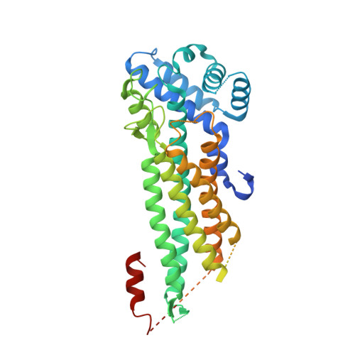
Adenylosuccinate lyase (PurB) catalyzes two distinct reactions in the purine nucleotide biosynthetic pathway using the same active site. The ability to recognize two different sets of substrates is of structural and evolutionary interest. In the present study, the crystal structure of PurB from the thermophilic bacterium Thermus thermophilus HB8 (TtPurB) was determined at a resolution of 2.38 Å by molecular replacement using a structure predicted by AlphaFold2 as a template. The asymmetric unit of the TtPurB crystal contained two TtPurB molecules, and some regions were disordered in the crystal structure. The disordered regions were the substrate-binding site and domain 3. TtPurB forms a homotetramer and the monomer is composed of three domains (domains 1, 2 and 3), which is a typical structure for the aspartase/fumarase superfamily. Molecular dynamics simulations with and without substrate/product were performed using a full-length model of TtPurB which was obtained before deletion of the disordered regions. The substrates and products were bound to the model structures during the MD simulations. The fluctuations of amino-acid residues were greater in the disordered regions and became smaller upon the binding of substrate or product. These results demonstrate that the full-length model obtained using AlphaFold2 can be used to generate the coordinates of disordered regions within the crystal structure.
- Faculty of Advanced Engineering, Chiba Institute of Technology, Narashino, Chiba 275-0016, Japan.
Organizational Affiliation: