Photocobilins integrate B12 and bilin photochemistry for enzyme control.
Zhang, S., Jeffreys, L.N., Poddar, H., Yu, Y., Liu, C., Patel, K., Johannissen, L.O., Zhu, L., Cliff, M.J., Yan, C., Schiro, G., Weik, M., Sakuma, M., Levy, C.W., Leys, D., Heyes, D.J., Scrutton, N.S.(2024) Nat Commun 15: 2740-2740
- PubMed: 38548733 Search on PubMedSearch on PubMed Central
- DOI: https://doi.org/10.1038/s41467-024-46995-1
- Primary Citation Related Structures:
8J2W, 8J2X, 8J2Y - PubMed Abstract:
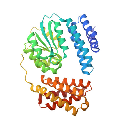
Photoreceptor proteins utilise chromophores to sense light and trigger a biological response. The discovery that adenosylcobalamin (or coenzyme B 12 ) can act as a light-sensing chromophore heralded a new field of B 12 -photobiology. Although microbial genome analysis indicates that photoactive B 12 -binding domains form part of more complex protein architectures, regulating a range of molecular-cellular functions in response to light, experimental evidence is lacking. Here we identify and characterise a sub-family of multi-centre photoreceptors, termed photocobilins, that use B 12 and biliverdin (BV) to sense light across the visible spectrum. Crystal structures reveal close juxtaposition of the B 12 and BV chromophores, an arrangement that facilitates optical coupling. Light-triggered conversion of the B 12 affects quaternary structure, in turn leading to light-activation of associated enzyme domains. The apparent widespread nature of photocobilins implies involvement in light regulation of a wider array of biochemical processes, and thus expands the scope for B 12 photobiology. Their characterisation provides inspiration for the design of broad-spectrum optogenetic tools and next generation bio-photocatalysts.
- Manchester Institute of Biotechnology and Department of Chemistry, The University of Manchester, 131 Princess Street, Manchester, M1 7DN, UK. shaowei.zhang@nudt.edu.cn.
Organizational Affiliation: