8J0H | pdb_00008j0h

Crystal structure of the fission yeast Rex1BD protein(C4H3.06)


Experimental Data Snapshot

  • Method: X-RAY DIFFRACTION
  • Resolution: 3.38 Å
  • R-Value Free: 
    0.296 (Depositor) 
  • R-Value Work: 
    0.242 (Depositor) 
  • R-Value Observed: 
    0.247 (Depositor) 

wwPDB Validation   3D Report Full Report


This is version 1.4 of the entry. See complete history


Literature

Rex1BD and the 14-3-3 protein control heterochromatin organization at tandem repeats by linking RNAi and HDAC.

Gao, J.Sun, W.Li, J.Ban, H.Zhang, T.Liao, J.Kim, N.Lee, S.H.Dong, Q.Madramootoo, R.Chen, Y.Li, F.

(2023) Proc Natl Acad Sci U S A 120: e2309359120-e2309359120

  • DOI: https://doi.org/10.1073/pnas.2309359120
  • Primary Citation of Related Structures:  
    8J0H

  • PubMed Abstract: 

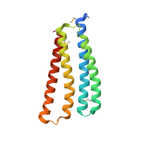
    Tandem DNA repeats are often organized into heterochromatin that is crucial for genome organization and stability. Recent studies revealed that individual repeats within tandem DNA repeats can behave very differently. How DNA repeats are assembled into distinct heterochromatin structures remains poorly understood. Here, we developed a genome-wide genetic screen using a reporter gene at different units in a repeat array. This screen led to identification of a conserved protein Rex1BD required for heterochromatin silencing. Our structural analysis revealed that Rex1BD forms a four-helix bundle structure with a distinct charged electrostatic surface. Mechanistically, Rex1BD facilitates the recruitment of Clr6 histone deacetylase (HDAC) by interacting with histones. Interestingly, Rex1BD also interacts with the 14-3-3 protein Rad25, which is responsible for recruiting the RITS (RNA-induced transcriptional silencing) complex to DNA repeats. Our results suggest that coordinated action of Rex1BD and Rad25 mediates formation of distinct heterochromatin structure at DNA repeats via linking RNAi and HDAC pathways.


  • Organizational Affiliation
    • Department of Biology, New York University, New York, NY 10003.

Macromolecules
Find similar proteins by:  (by identity cutoff)  |  3D Structure
Entity ID: 1
MoleculeChains Sequence LengthOrganismDetailsImage
Uncharacterized protein C4H3.06
A, B, C, D, E
A, B, C, D, E, F, G, H
142Schizosaccharomyces pombe 972h-Mutation(s): 0 
Gene Names: SPAC4H3.06
UniProt
Find proteins for Q10214 (Schizosaccharomyces pombe (strain 972 / ATCC 24843))
Explore Q10214 
Go to UniProtKB:  Q10214
Entity Groups  
Sequence Clusters30% Identity50% Identity70% Identity90% Identity95% Identity100% Identity
UniProt GroupQ10214
Sequence Annotations
Expand
  • Reference Sequence
Small Molecules
Modified Residues  1 Unique
IDChains TypeFormula2D DiagramParent
MSE
Query on MSE
A, B, C, D, E
A, B, C, D, E, F, G, H
L-PEPTIDE LINKINGC5 H11 N O2 SeMET
Experimental Data & Validation

Experimental Data

  • Method: X-RAY DIFFRACTION
  • Resolution: 3.38 Å
  • R-Value Free:  0.296 (Depositor) 
  • R-Value Work:  0.242 (Depositor) 
  • R-Value Observed: 0.247 (Depositor) 
Space Group: P 21 21 2
Unit Cell:
Length ( Å )Angle ( ˚ )
a = 159.014α = 90
b = 138.481β = 90
c = 50.897γ = 90
Software Package:
Software NamePurpose
PHENIXrefinement
XDSdata scaling
XDSdata reduction
PHASESphasing

Structure Validation

View Full Validation Report


Entry History & Funding Information

Deposition Data


Funding OrganizationLocationGrant Number
Chinese Academy of SciencesChinaXDB37010303

Revision History  (Full details and data files)

  • Version 1.0: 2023-11-01
    Type: Initial release
  • Version 1.1: 2023-11-15
    Changes: Data collection
  • Version 1.2: 2024-05-29
    Changes: Database references
  • Version 1.3: 2024-08-14
    Changes: Database references
  • Version 1.4: 2024-10-16
    Changes: Structure summary