8ITG | pdb_00008itg

Crystal structure of lasso peptide epimerase MslH in complexed with precursor peptide variant MslAW21G


Experimental Data Snapshot

  • Method: X-RAY DIFFRACTION
  • Resolution: 2.25 Å
  • R-Value Free: 
    0.207 (Depositor), 0.208 (DCC) 
  • R-Value Work: 
    0.182 (Depositor), 0.184 (DCC) 
  • R-Value Observed: 
    0.184 (Depositor) 

Starting Model: experimental
View more details

wwPDB Validation 3D Report Full Report

Validation slider image for 8ITG

This is version 1.3 of the entry. See complete history

Literature

Structure of lasso peptide epimerase MslH reveals metal-dependent acid/base catalytic mechanism.

Nakashima, Y.Kawakami, A.Ogasawara, Y.Maeki, M.Tokeshi, M.Dairi, T.Morita, H.

(2023) Nat Commun 14: 4752-4752

  • DOI: https://doi.org/10.1038/s41467-023-40232-x
  • Primary Citation Related Structures: 
    8GQ9, 8GQA, 8GQB, 8ITG, 8ITH

  • PubMed Abstract: 

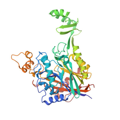
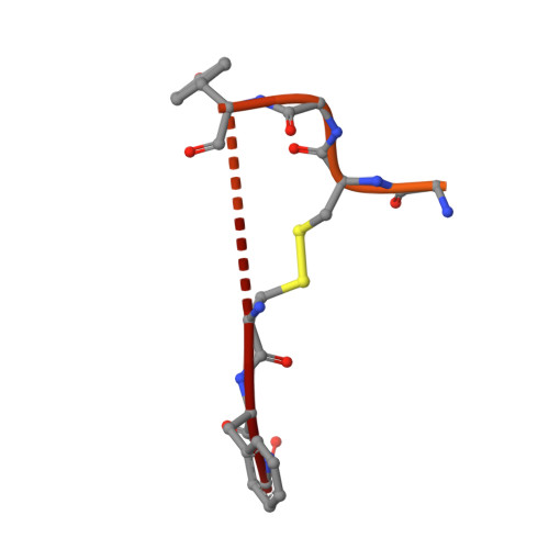
    The lasso peptide MS-271 is a ribosomally synthesized and post-translationally modified peptide (RiPP) consisting of 21 amino acids with D-tryptophan at the C-terminus, and is derived from the precursor peptide MslA. MslH, encoded in the MS-271 biosynthetic gene cluster (msl), catalyzes the epimerization at the Cα center of the MslA C-terminal Trp21, leading to epi-MslA. The detailed catalytic process, including the catalytic site and cofactors, has remained enigmatic. Herein, based on X-ray crystallographic studies in association with MslA core peptide analogues, we show that MslH is a metallo-dependent peptide epimerase with a calcineurin-like fold. The crystal structure analysis, followed by site-directed mutagenesis, docking simulation, and ICP-MS studies demonstrate that MslH employs acid/base chemistry to facilitate the reversible epimerization of the C-terminal Trp21 of MslA, by utilizing two pairs of His/Asp catalytic residues that are electrostatically tethered to a six-coordination motif with a Ca(II) ion via water molecules.


  • Organizational Affiliation
    • Institute of Natural Medicine, University of Toyama, 2630-Sugitani, Toyama, 930-0194, Japan.

Macromolecule Content 

  • Total Structure Weight: 51.92 kDa 
  • Atom Count: 3,627 
  • Modeled Residue Count: 446 
  • Deposited Residue Count: 482 
  • Unique protein chains: 2

Macromolecules

Find similar proteins by:|  3D Structure
Entity ID: 1
MoleculeChains  Sequence LengthOrganismDetailsImage
Poly-gamma-glutamate synthesis protein (Capsule biosynthesis protein)440Streptomyces griseorubiginosusMutation(s): 0 
Gene Names: EV578_104528
Find similar proteins by:|  3D Structure
Entity ID: 2
MoleculeChains  Sequence LengthOrganismDetailsImage
Tricyclic peptide MS-27142Streptomyces griseorubiginosusMutation(s): 0 
Gene Names: DWG14_02265

Small Molecules

Experimental Data & Validation

Experimental Data

  • Method: X-RAY DIFFRACTION
  • Resolution: 2.25 Å
  • R-Value Free:  0.207 (Depositor), 0.208 (DCC) 
  • R-Value Work:  0.182 (Depositor), 0.184 (DCC) 
  • R-Value Observed: 0.184 (Depositor) 
Space Group: I 4 2 2
Unit Cell:
Length ( Å )Angle ( ˚ )
a = 127.232α = 90
b = 127.232β = 90
c = 170.611γ = 90
Software Package:
Software NamePurpose
PHENIXrefinement
XDSdata reduction
Aimlessdata scaling
PHASERphasing

Structure Validation

View Full Validation Report


Entry History 

& Funding Information

Deposition Data


Funding OrganizationLocationGrant Number
Japan Society for the Promotion of Science (JSPS)JapanJP22H02777
Japan Society for the Promotion of Science (JSPS)JapanJP22K15303

Revision History  (Full details and data files)

  • Version 1.0: 2023-06-21
    Type: Initial release
  • Version 1.1: 2023-08-16
    Changes: Data collection, Database references
  • Version 1.2: 2023-08-23
    Changes: Database references
  • Version 1.3: 2024-10-23
    Changes: Structure summary