8IT0 | pdb_00008it0

Cryo-EM structure of Crt-SPARTA-gRNA-tDNA dimer (conformation-2)


Experimental Data Snapshot

  • Method: ELECTRON MICROSCOPY
  • Resolution: 3.50 Å
  • Aggregation State: PARTICLE 
  • Reconstruction Method: SINGLE PARTICLE 

wwPDB Validation   3D Report Full Report


This is version 1.2 of the entry. See complete history


Literature

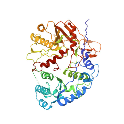
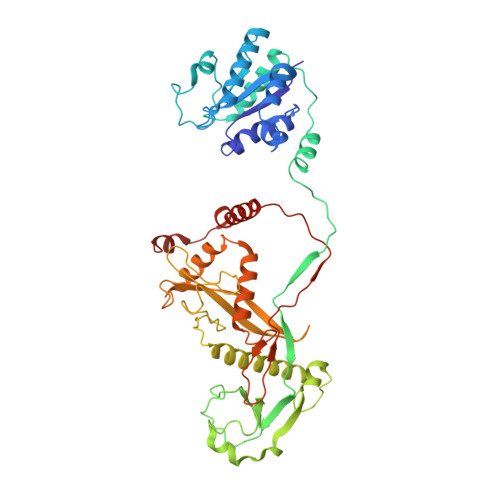
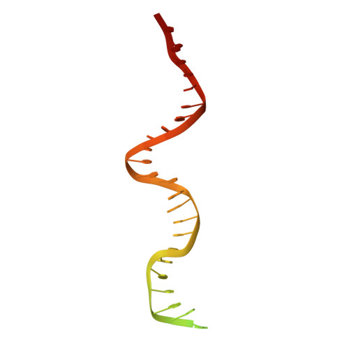
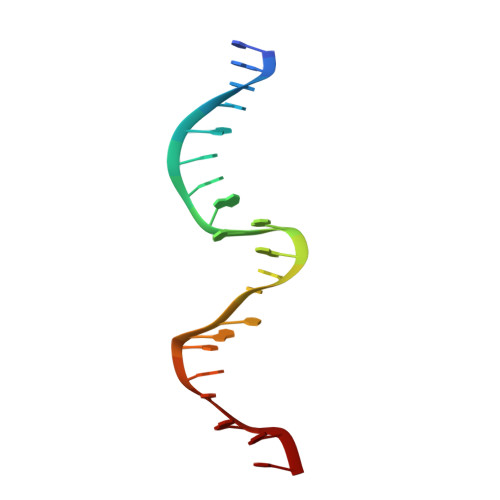
Nucleic-acid-triggered NADase activation of a short prokaryotic Argonaute.

Gao, X.Shang, K.Zhu, K.Wang, L.Mu, Z.Fu, X.Yu, X.Qin, B.Zhu, H.Ding, W.Cui, S.

(2024) Nature 625: 822-831

  • DOI: https://doi.org/10.1038/s41586-023-06665-6
  • Primary Citation of Related Structures:  
    8ISY, 8ISZ, 8IT0, 8IT1, 8K9G

  • PubMed Abstract: 

    Argonaute (Ago) proteins mediate RNA- or DNA-guided inhibition of nucleic acids 1,2 . Although the mechanisms used by eukaryotic Ago proteins and long prokaryotic Ago proteins (pAgos) are known, that used by short pAgos remains elusive. Here we determined the cryo-electron microscopy structures of a short pAgo and the associated TIR-APAZ proteins (SPARTA) from Crenotalea thermophila (Crt): a free-state Crt-SPARTA; a guide RNA-target DNA-loaded Crt-SPARTA; two Crt-SPARTA dimers with distinct TIR organization; and a Crt-SPARTA tetramer. These structures reveal that Crt-SPARTA is composed of a bilobal-fold Ago lobe that connects with a TIR lobe. Whereas the Crt-Ago contains a MID and a PIWI domain, Crt-TIR-APAZ has a TIR domain, an N-like domain, a linker domain and a trigger domain. The bound RNA-DNA duplex adopts a B-form conformation that is recognized by base-specific contacts. Nucleic acid binding causes conformational changes because the trigger domain acts as a 'roadblock' that prevents the guide RNA 5' ends and the target DNA 3' ends from reaching their canonical pockets; this disorders the MID domain and promotes Crt-SPARTA dimerization. Two RNA-DNA-loaded Crt-SPARTA dimers form a tetramer through their TIR domains. Four Crt-TIR domains assemble into two parallel head-to-tail-organized TIR dimers, indicating an NADase-active conformation, which is supported by our mutagenesis study. Our results reveal the structural basis of short-pAgo-mediated defence against invading nucleic acids, and provide insights for optimizing the detection of SPARTA-based programmable DNA sequences.


  • Organizational Affiliation
    • NHC Key Laboratory of Systems Biology of Pathogens, Institute of Pathogen Biology, Chinese Academy of Medical Sciences and Peking Union Medical College, Beijing, China.

Macromolecules

Find similar proteins by:  (by identity cutoff)  |  3D Structure
Entity ID: 1
MoleculeChains Sequence LengthOrganismDetailsImage
Piwi domain-containing proteinA,
C [auth E]
507Thermoflavifilum thermophilumMutation(s): 0 
Gene Names: SAMN05660895_1671
UniProt
Find proteins for A0A1I7NFD7 (Thermoflavifilum thermophilum)
Explore A0A1I7NFD7 
Go to UniProtKB:  A0A1I7NFD7
Entity Groups  
Sequence Clusters30% Identity50% Identity70% Identity90% Identity95% Identity100% Identity
UniProt GroupA0A1I7NFD7
Sequence Annotations
Expand
  • Reference Sequence
Find similar proteins by:  (by identity cutoff)  |  3D Structure
Entity ID: 2
MoleculeChains Sequence LengthOrganismDetailsImage
TIR domain-containing proteinB,
D [auth F]
450Thermoflavifilum thermophilumMutation(s): 0 
Gene Names: SAMN05660895_1670
UniProt
Find proteins for A0A1I7NFG5 (Thermoflavifilum thermophilum)
Explore A0A1I7NFG5 
Go to UniProtKB:  A0A1I7NFG5
Entity Groups  
Sequence Clusters30% Identity50% Identity70% Identity90% Identity95% Identity100% Identity
UniProt GroupA0A1I7NFG5
Sequence Annotations
Expand
  • Reference Sequence
Find similar nucleic acids by:  (by identity cutoff)  |  3D Structure
Entity ID: 3
MoleculeChains LengthOrganismImage
DNA (45-mer)E [auth D],
G [auth H]
45synthetic construct
Sequence Annotations
Expand
  • Reference Sequence

Find similar nucleic acids by:  Sequence   |   3D Structure  

Entity ID: 4
MoleculeChains LengthOrganismImage
RNA (5'-R(P*UP*GP*AP*GP*GP*UP*AP*GP*UP*AP*GP*GP*UP*UP*GP*UP*AP*UP*AP*GP*U)-3')F [auth C],
H [auth G]
21synthetic construct
Sequence Annotations
Expand
  • Reference Sequence
Experimental Data & Validation

Experimental Data

  • Method: ELECTRON MICROSCOPY
  • Resolution: 3.50 Å
  • Aggregation State: PARTICLE 
  • Reconstruction Method: SINGLE PARTICLE 
EM Software:
TaskSoftware PackageVersion
MODEL REFINEMENTPHENIX1.20.1
RECONSTRUCTIONcryoSPARC4.2

Structure Validation

View Full Validation Report


Entry History & Funding Information

Deposition Data


Funding OrganizationLocationGrant Number
Not funded--

Revision History  (Full details and data files)

  • Version 1.0: 2023-10-18
    Type: Initial release
  • Version 1.1: 2023-11-08
    Changes: Data collection, Structure summary
  • Version 1.2: 2024-02-07
    Changes: Database references