8ISN

HLA-A24 in complex with modified 9mer WT1 peptide


Experimental Data Snapshot

  • Method: X-RAY DIFFRACTION
  • Resolution: 2.48 Å
  • R-Value Free: 0.258 
  • R-Value Work: 0.222 
  • R-Value Observed: 0.224 

wwPDB Validation   3D Report Full Report


This is version 1.2 of the entry. See complete history


Literature

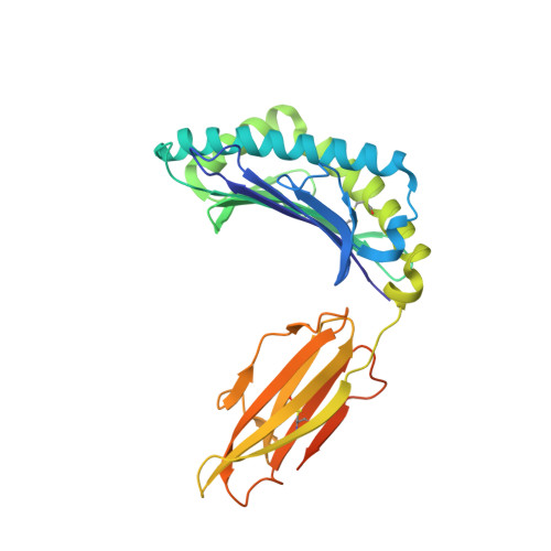
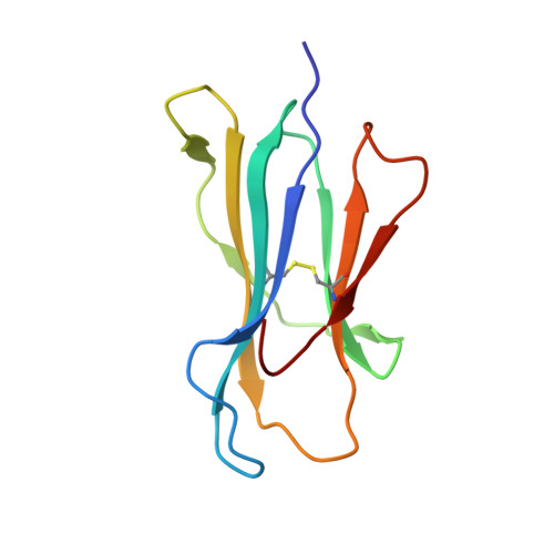
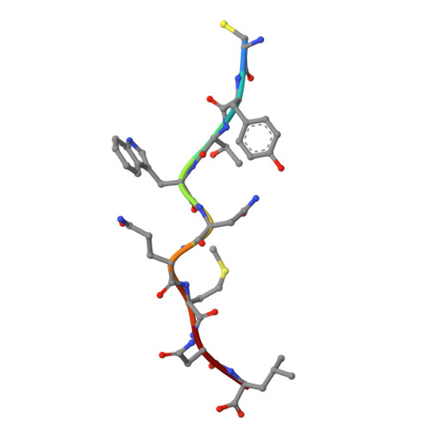
Elucidation of binding mechanism, affinity, and complex structure between mWT1 tumor-associated antigen peptide and HLA-A*24:02.

Bekker, G.J.Numoto, N.Kawasaki, M.Hayashi, T.Yabuno, S.Kozono, Y.Shimizu, T.Kozono, H.Ito, N.Oda, M.Kamiya, N.

(2023) Protein Sci 32: e4775-e4775

  • DOI: https://doi.org/10.1002/pro.4775
  • Primary Citation of Related Structures:  
    8ISN

  • PubMed Abstract: 

    We have applied our advanced computational and experimental methodologies to investigate the complex structure and binding mechanism of a modified Wilms' Tumor 1 (mWT1) protein epitope to the understudied Asian-dominant allele HLA-A*24:02 (HLA-A24) in aqueous solution. We have applied our developed multicanonical molecular dynamics (McMD)-based dynamic docking method to analyze the binding pathway and mechanism, which we verified by comparing the highest probability structures from simulation with our experimentally solved x-ray crystal structure. Subsequent path sampling MD simulations elucidated the atomic details of the binding process and indicated that first an encounter complex is formed between the N-terminal's positive charge of the 9-residue mWT1 fragment peptide and a cluster of negative residues on the surface of HLA-A24, with the major histocompatibility complex (MHC) molecule preferring a predominantly closed conformation. The peptide first binds to this closed MHC conformation, forming an encounter complex, after which the binding site opens due to increased entropy of the binding site, allowing the peptide to bind to form the native complex structure. Further sequence and structure analyses also suggest that although the peptide loading complex would help with stabilizing the MHC molecule, the binding depends in a large part on the intrinsic affinity between the MHC molecule and the antigen peptide. Finally, our computational tools and analyses can be of great benefit to study the binding mechanism of different MHC types to their antigens, where it could also be useful in the development of higher affinity variant peptides and for personalized medicine.


  • Organizational Affiliation

    Institute for Protein Research, Osaka University, Suita, Osaka, Japan.


Macromolecules
Find similar proteins by:  (by identity cutoff)  |  3D Structure
Entity ID: 1
MoleculeChains Sequence LengthOrganismDetailsImage
MHC class I antigen
A, D
305Homo sapiensMutation(s): 0 
Gene Names: HLA-A
UniProt
Find proteins for D9UAY1 (Homo sapiens)
Explore D9UAY1 
Go to UniProtKB:  D9UAY1
Entity Groups  
Sequence Clusters30% Identity50% Identity70% Identity90% Identity95% Identity100% Identity
UniProt GroupD9UAY1
Sequence Annotations
Expand
  • Reference Sequence
Find similar proteins by:  (by identity cutoff)  |  3D Structure
Entity ID: 2
MoleculeChains Sequence LengthOrganismDetailsImage
Beta-2-microglobulin
B, E
99Homo sapiensMutation(s): 0 
Gene Names: B2M
UniProt & NIH Common Fund Data Resources
Find proteins for P61769 (Homo sapiens)
Explore P61769 
Go to UniProtKB:  P61769
PHAROS:  P61769
GTEx:  ENSG00000166710 
Entity Groups  
Sequence Clusters30% Identity50% Identity70% Identity90% Identity95% Identity100% Identity
UniProt GroupP61769
Sequence Annotations
Expand
  • Reference Sequence

Find similar proteins by:  Sequence   |   3D Structure  

Entity ID: 3
MoleculeChains Sequence LengthOrganismDetailsImage
CYS-TYR-THR-TRP-ASN-GLN-MET-ASN-LEU
C, F
9Homo sapiensMutation(s): 0 
UniProt & NIH Common Fund Data Resources
Find proteins for P19544 (Homo sapiens)
Explore P19544 
Go to UniProtKB:  P19544
PHAROS:  P19544
GTEx:  ENSG00000184937 
Entity Groups  
UniProt GroupP19544
Sequence Annotations
Expand
  • Reference Sequence
Oligosaccharides

Help

Entity ID: 4
MoleculeChains Length2D Diagram Glycosylation3D Interactions
alpha-D-mannopyranose-(1-3)-beta-D-mannopyranose-(1-4)-2-acetamido-2-deoxy-beta-D-glucopyranose-(1-4)-2-acetamido-2-deoxy-beta-D-glucopyranose
G
4N-Glycosylation
Glycosylation Resources
GlyTouCan:  G81315DD
GlyCosmos:  G81315DD
GlyGen:  G81315DD
Experimental Data & Validation

Experimental Data

  • Method: X-RAY DIFFRACTION
  • Resolution: 2.48 Å
  • R-Value Free: 0.258 
  • R-Value Work: 0.222 
  • R-Value Observed: 0.224 
  • Space Group: F 2 2 2
Unit Cell:
Length ( Å )Angle ( ˚ )
a = 161.4α = 90
b = 166.13β = 90
c = 180.53γ = 90
Software Package:
Software NamePurpose
PHENIXrefinement
XDSdata reduction
XSCALEdata scaling
PHASERphasing

Structure Validation

View Full Validation Report


Entry History & Funding Information

Deposition Data


Funding OrganizationLocationGrant Number
Japan Society for the Promotion of Science (JSPS)JapanJP20H03229
Japan Agency for Medical Research and Development (AMED)JapanJP20fk0108471
Japan Agency for Medical Research and Development (AMED)JapanJP21zf0127004

Revision History  (Full details and data files)

  • Version 1.0: 2023-09-13
    Type: Initial release
  • Version 1.1: 2023-10-11
    Changes: Database references
  • Version 1.2: 2023-10-25
    Changes: Database references