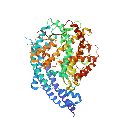
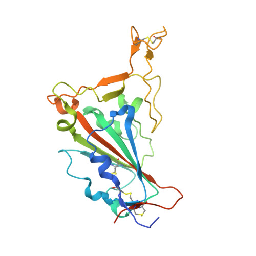
In late 2022, various Omicron subvariants emerged and cocirculated worldwide. These variants convergently acquired amino acid substitutions at critical residues in the spike protein, including residues R346, K444, L452, N460, and F486. Here, we characterize the convergent evolution of Omicron subvariants and the properties of one recent lineage of concern, BQ.1.1. Our phylogenetic analysis suggests that these five substitutions are recurrently acquired, particularly in younger Omicron lineages. Epidemic dynamics modelling suggests that the five substitutions increase viral fitness, and a large proportion of the fitness variation within Omicron lineages can be explained by these substitutions. Compared to BA.5, BQ.1.1 evades breakthrough BA.2 and BA.5 infection sera more efficiently, as demonstrated by neutralization assays. The pathogenicity of BQ.1.1 in hamsters is lower than that of BA.5. Our multiscale investigations illuminate the evolutionary rules governing the convergent evolution for known Omicron lineages as of 2022.
Organizational Affiliation:
Division of Systems Virology, Department of Microbiology and Immunology, The Institute of Medical Science, The University of Tokyo, Tokyo, Japan.
Department of Microbiology and Immunology, Faculty of Medicine, Hokkaido University, Sapporo, Japan.
Graduate School of Medicine, The University of Tokyo, Tokyo, Japan.
Division of Molecular Pathobiology, International Institute for Zoonosis Control, Hokkaido University, Sapporo, Japan.
Department of Biomolecular Sciences, Weizmann Institute of Science, Rehovot, Israel.
First Medical Faculty at Biocev, Charles University, Vestec-Prague, Czechia.
Laboratory of Medical Virology, Institute for Life and Medical Sciences, Kyoto University, Kyoto, Japan.
Center for iPS Cell Research and Application (CiRA), Kyoto University, Kyoto, Japan.
Department of Cancer Pathology, Faculty of Medicine, Hokkaido University, Sapporo, Japan.
Institute for Chemical Reaction Design and Discovery (WPI-ICReDD), Hokkaido University, Sapporo, Japan.
Medical Research Council-University of Glasgow Centre for Virus Research, Glasgow, UK.
Division of Risk Analysis and Management, International Institute for Zoonosis Control, Hokkaido University, Sapporo, Japan.
Division of Molecular Virology and Genetics, Joint Research Center for Human Retrovirus infection, Kumamoto University, Kumamoto, Japan.
Department of Clinical Pathology, Faculty of Medicine, Suez Canal University, Ismailia, Egypt.
Department of Veterinary Science, Faculty of Agriculture, University of Miyazaki, Miyazaki, Japan.
Graduate School of Medicine and Veterinary Medicine, University of Miyazaki, Miyazaki, Japan.
Department of Medicinal Sciences, Graduate School of Pharmaceutical Sciences, Kyushu University, Fukuoka, Japan.
Institute for Genetic Medicine, Hokkaido University, Sapporo, Japan.
Division of International Research Promotion, International Institute for Zoonosis Control, Hokkaido University, Sapporo, Japan.
One Health Research Center, Hokkaido University, Sapporo, Japan.
Institute for Vaccine Research and Development: HU-IVReD, Hokkaido University, Sapporo, Japan.
Tokyo Metropolitan Institute of Public Health, Tokyo, Japan.
HiLung, Inc, Kyoto, Japan.
Interpark Kuramochi Clinic, Utsunomiya, Japan.
Department of Global Health Promotion, Tokyo Medical and Dental University, Tokyo, Japan.
Center for Animal Disease Control, University of Miyazaki, Miyazaki, Japan.
International Collaboration Unit, International Institute for Zoonosis Control, Hokkaido University, Sapporo, Japan.
AMED-CREST, Japan Agency for Medical Research and Development (AMED), Tokyo, Japan.
Laboratory of Medical Virology, Institute for Life and Medical Sciences, Kyoto University, Kyoto, Japan. hashiguchi.takao.1a@kyoto-u.ac.jp.
Department of Cancer Pathology, Faculty of Medicine, Hokkaido University, Sapporo, Japan. tanaka@med.hokudai.ac.jp.
Institute for Chemical Reaction Design and Discovery (WPI-ICReDD), Hokkaido University, Sapporo, Japan. tanaka@med.hokudai.ac.jp.
Department of Microbiology and Immunology, Faculty of Medicine, Hokkaido University, Sapporo, Japan. fukut@pop.med.hokudai.ac.jp.
AMED-CREST, Japan Agency for Medical Research and Development (AMED), Tokyo, Japan. fukut@pop.med.hokudai.ac.jp.
Laboratory of Virus Control, Research Institute for Microbial Diseases, Osaka University, Suita, Japan. fukut@pop.med.hokudai.ac.jp.
Division of Molecular Virology and Genetics, Joint Research Center for Human Retrovirus infection, Kumamoto University, Kumamoto, Japan. ikedat@kumamoto-u.ac.jp.
Division of Systems Virology, Department of Microbiology and Immunology, The Institute of Medical Science, The University of Tokyo, Tokyo, Japan. KeiSato@g.ecc.u-tokyo.ac.jp.
Graduate School of Medicine, The University of Tokyo, Tokyo, Japan. KeiSato@g.ecc.u-tokyo.ac.jp.
International Research Center for Infectious Diseases, The Institute of Medical Science, The University of Tokyo, Tokyo, Japan. KeiSato@g.ecc.u-tokyo.ac.jp.
International Vaccine Design Center, The Institute of Medical Science, The University of Tokyo, Tokyo, Japan. KeiSato@g.ecc.u-tokyo.ac.jp.
Graduate School of Frontier Sciences, The University of Tokyo, Kashiwa, Japan. KeiSato@g.ecc.u-tokyo.ac.jp.
Collaboration Unit for Infection, Joint Research Center for Human Retrovirus infection, Kumamoto University, Kumamoto, Japan. KeiSato@g.ecc.u-tokyo.ac.jp.
CREST, Japan Science and Technology Agency, Kawaguchi, Japan. KeiSato@g.ecc.u-tokyo.ac.jp.