8IDN

Cryo-EM structure of RBD/E77-Fab complex


Experimental Data Snapshot

  • Method: ELECTRON MICROSCOPY
  • Resolution: 3.35 Å
  • Aggregation State: PARTICLE 
  • Reconstruction Method: SINGLE PARTICLE 

wwPDB Validation   3D Report Full Report


This is version 1.1 of the entry. See complete history


Literature

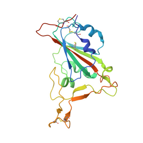
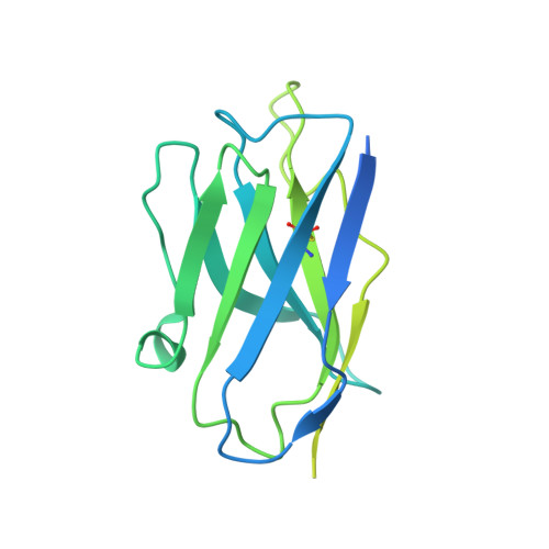
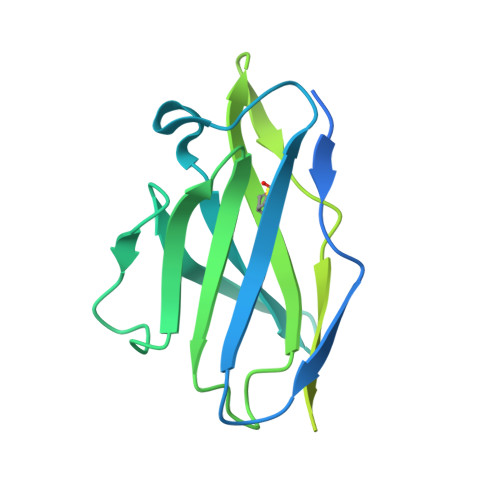
The structure of the RBD-E77 Fab complex reveals neutralization and immune escape of SARS-CoV-2.

Zhang, Z.Li, X.Xue, Y.Yang, B.Jia, Y.Liu, S.Lu, D.

(2023) Acta Crystallogr D Struct Biol 79: 746-757

  • DOI: https://doi.org/10.1107/S2059798323005041
  • Primary Citation of Related Structures:  
    8IDN

  • PubMed Abstract: 

    The spike protein (S) of SARS-CoV-2 is the major target of neutralizing antibodies and vaccines. Antibodies that target the receptor-binding domain (RBD) of S have high potency in preventing viral infection. The ongoing evolution of SARS-CoV-2, especially mutations occurring in the RBD of new variants, has severely challenged the development of neutralizing antibodies and vaccines. Here, a murine monoclonal antibody (mAb) designated E77 is reported which engages the prototype RBD with high affinity and potently neutralizes SARS-CoV-2 pseudoviruses. However, the capability of E77 to bind RBDs vanishes upon encountering variants of concern (VOCs) which carry the N501Y mutation, such as Alpha, Beta, Gamma and Omicron, in contrast to its performance with the Delta variant. To explain the discrepancy, cryo-electron microscopy was used to analyze the structure of an RBD-E77 Fab complex, which reveals that the binding site of E77 on RBD belongs to the RBD-1 epitope, which largely overlaps with the binding site of human angiotensin-converting enzyme 2 (hACE2). Both the heavy chain and the light chain of E77 interact extensively with RBD and contribute to the strong binding of RBD. E77 employs CDRL1 to engage Asn501 of RBD and the Asn-to-Tyr mutation could generate steric hindrance, abolishing the binding. In sum, the data provide the landscape for an in-depth understanding of immune escape of VOCs and rational antibody engineering against emerging variants of SARS-CoV-2.


  • Organizational Affiliation

    College of Life Sciences, Shanxi Agricultural University, Taiyuan 030031, People's Republic of China.


Macromolecules
Find similar proteins by:  (by identity cutoff)  |  3D Structure
Entity ID: 1
MoleculeChains Sequence LengthOrganismDetailsImage
Spike protein S1206Severe acute respiratory syndrome coronavirus 2Mutation(s): 0 
Gene Names: S2
UniProt
Find proteins for P0DTC2 (Severe acute respiratory syndrome coronavirus 2)
Explore P0DTC2 
Go to UniProtKB:  P0DTC2
Entity Groups  
Sequence Clusters30% Identity50% Identity70% Identity90% Identity95% Identity100% Identity
UniProt GroupP0DTC2
Sequence Annotations
Expand
  • Reference Sequence
Find similar proteins by:  (by identity cutoff)  |  3D Structure
Entity ID: 2
MoleculeChains Sequence LengthOrganismDetailsImage
E77 Fab heavy chainB [auth H]239Mus musculusMutation(s): 0 
Entity Groups  
Sequence Clusters30% Identity50% Identity70% Identity90% Identity95% Identity100% Identity
Sequence Annotations
Expand
  • Reference Sequence
Find similar proteins by:  (by identity cutoff)  |  3D Structure
Entity ID: 3
MoleculeChains Sequence LengthOrganismDetailsImage
E77 Fab light chainC [auth L]233Mus musculusMutation(s): 0 
Entity Groups  
Sequence Clusters30% Identity50% Identity70% Identity90% Identity95% Identity100% Identity
Sequence Annotations
Expand
  • Reference Sequence
Oligosaccharides

Help

Entity ID: 4
MoleculeChains Length2D Diagram Glycosylation3D Interactions
alpha-D-mannopyranose-(1-3)-beta-D-mannopyranose-(1-4)-2-acetamido-2-deoxy-beta-D-glucopyranose-(1-4)-2-acetamido-2-deoxy-beta-D-glucopyranoseD [auth B]4N-Glycosylation
Glycosylation Resources
GlyTouCan:  G81315DD
GlyCosmos:  G81315DD
GlyGen:  G81315DD
Experimental Data & Validation

Experimental Data

  • Method: ELECTRON MICROSCOPY
  • Resolution: 3.35 Å
  • Aggregation State: PARTICLE 
  • Reconstruction Method: SINGLE PARTICLE 

Structure Validation

View Full Validation Report


Entry History & Funding Information

Deposition Data


Funding OrganizationLocationGrant Number
Ministry of Science and Technology (MoST, China)China2021YFC2301400

Revision History  (Full details and data files)

  • Version 1.0: 2023-06-21
    Type: Initial release
  • Version 1.1: 2023-08-16
    Changes: Data collection, Database references