8IDA | pdb_00008ida

Overall structure of the LAT1-4F2hc bound with tyrosine


Experimental Data Snapshot

  • Method: ELECTRON MICROSCOPY
  • Resolution: 3.20 Å
  • Aggregation State: PARTICLE 
  • Reconstruction Method: SINGLE PARTICLE 

wwPDB Validation   3D Report Full Report


This is version 2.2 of the entry. See complete history


Literature

Structural insights into the substrate transport mechanism of the amino acid transporter complex.

Yang, H.Shi, T.Dong, J.Zhang, T.Li, Y.Guo, Y.Yuan, Y.Yang, L.Dong, J.T.Yan, R.

(2025) J Biological Chem 301: 110569-110569

  • DOI: https://doi.org/10.1016/j.jbc.2025.110569
  • Primary Citation of Related Structures:  
    8IDA, 8J8L, 8J8M, 8X0W

  • PubMed Abstract: 

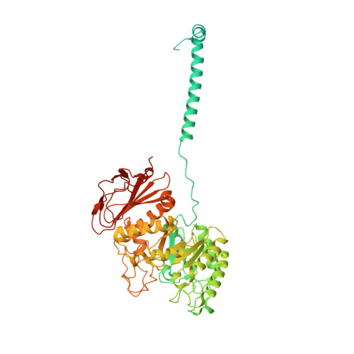
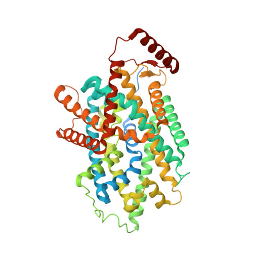
    The L -type amino acid transporter 1 (LAT1), in complex with its ancillary protein 4F2hc, mediates the sodium-independent antiport of large neutral amino acids across the plasma membrane. LAT1 preferentially transports substrates, such as L -leucine, L -tyrosine, and L -tryptophan, thyroid hormones, and drugs like 3,4-dihydroxyphenylalanine. Its pivotal role in cancer development and progression has established LAT1 as a promising therapeutic target. While prior studies have resolved the LAT1-4F2hc architecture and inhibitor interactions, the molecular basis of LAT1 substrate selectivity remains elusive. Here, we present the cryo-EM structures of LAT1-4F2hc bound to L -tyrosine, L -tryptophan, L -leucine, and 3,4-dihydroxyphenylalanine, revealing distinct substrate binding modes. Comparative structural analysis highlights differences between LAT1 and LAT2 in substrate coordination, driven by key residues near the binding pocket that influence transport efficiency. These findings advance our mechanistic understanding of the LAT1-4F2hc complex and provide valuable insights for structure-based drug design targeting LAT1.


  • Organizational Affiliation
    • Department of Biochemistry, SUSTech Homeostatic Medicine Institute, School of Medicine, Key University Laboratory of Metabolism and Health of Guangdong, Institute for Biological Electron Microscopy, Southern University of Science and Technology, Shenzhen, Guangdong, China.

Macromolecules
Find similar proteins by:  (by identity cutoff)  |  3D Structure
Entity ID: 1
MoleculeChains Sequence LengthOrganismDetailsImage
4F2 cell-surface antigen heavy chain647Homo sapiensMutation(s): 0 
Gene Names: SLC3A2MDU1
Membrane Entity: Yes 
UniProt & NIH Common Fund Data Resources
Find proteins for P08195 (Homo sapiens)
Explore P08195 
Go to UniProtKB:  P08195
PHAROS:  P08195
GTEx:  ENSG00000168003 
Entity Groups  
Sequence Clusters30% Identity50% Identity70% Identity90% Identity95% Identity100% Identity
UniProt GroupP08195
Glycosylation
Glycosylation Sites: 4Go to GlyGen: P08195-1
Sequence Annotations
Expand
  • Reference Sequence
Find similar proteins by:  (by identity cutoff)  |  3D Structure
Entity ID: 2
MoleculeChains Sequence LengthOrganismDetailsImage
Large neutral amino acids transporter small subunit 1527Homo sapiensMutation(s): 0 
Gene Names: SLC7A5CD98LCLAT1MPE16
Membrane Entity: Yes 
UniProt & NIH Common Fund Data Resources
Find proteins for Q01650 (Homo sapiens)
Explore Q01650 
Go to UniProtKB:  Q01650
PHAROS:  Q01650
GTEx:  ENSG00000103257 
Entity Groups  
Sequence Clusters30% Identity50% Identity70% Identity90% Identity95% Identity100% Identity
UniProt GroupQ01650
Sequence Annotations
Expand
  • Reference Sequence
Oligosaccharides

Help

Entity ID: 3
MoleculeChains Length2D Diagram Glycosylation3D Interactions
2-acetamido-2-deoxy-beta-D-glucopyranose-(1-4)-2-acetamido-2-deoxy-beta-D-glucopyranoseC,
D,
E [auth F],
F [auth E]
2N-Glycosylation
Glycosylation Resources
GlyTouCan:  G42666HT
GlyCosmos:  G42666HT
GlyGen:  G42666HT
Small Molecules
Ligands 1 Unique
IDChains Name / Formula / InChI Key2D Diagram3D Interactions
TYR
Query on TYR

Download Ideal Coordinates CCD File 
G [auth B]TYROSINE
C9 H11 N O3
OUYCCCASQSFEME-QMMMGPOBSA-N
Experimental Data & Validation

Experimental Data

  • Method: ELECTRON MICROSCOPY
  • Resolution: 3.20 Å
  • Aggregation State: PARTICLE 
  • Reconstruction Method: SINGLE PARTICLE 

Structure Validation

View Full Validation Report


Entry History & Funding Information

Deposition Data


Funding OrganizationLocationGrant Number
National Natural Science Foundation of China (NSFC)China--

Revision History  (Full details and data files)

  • Version 1.0: 2024-07-10
    Type: Initial release
  • Version 2.0: 2024-07-17
    Type: Coordinate replacement
    Reason: Atoms with unrealistic or zero occupancies
    Changes: Atomic model, Author supporting evidence, Data collection, Derived calculations, Non-polymer description, Structure summary
  • Version 2.1: 2024-10-30
    Changes: Data collection, Structure summary
  • Version 2.2: 2025-11-26
    Changes: Data collection, Database references