8IAU

Crystal structure of Streptococcus pneumoniae pyruvate kinase in complex with oxalate and fructose 1,6-bisphosphate


Experimental Data Snapshot

  • Method: X-RAY DIFFRACTION
  • Resolution: 2.00 Å
  • R-Value Free: 0.246 
  • R-Value Work: 0.200 
  • R-Value Observed: 0.202 

wwPDB Validation   3D Report Full Report


Ligand Structure Quality Assessment 


This is version 1.2 of the entry. See complete history


Literature

Functional and structural characterization of Streptococcus pneumoniae pyruvate kinase involved in fosfomycin resistance.

Taguchi, A.Nakashima, R.Nishino, K.

(2023) J Biol Chem 299: 104892-104892

  • DOI: https://doi.org/10.1016/j.jbc.2023.104892
  • Primary Citation of Related Structures:  
    8IAS, 8IAT, 8IAU, 8IAV, 8IAW, 8IAX

  • PubMed Abstract: 

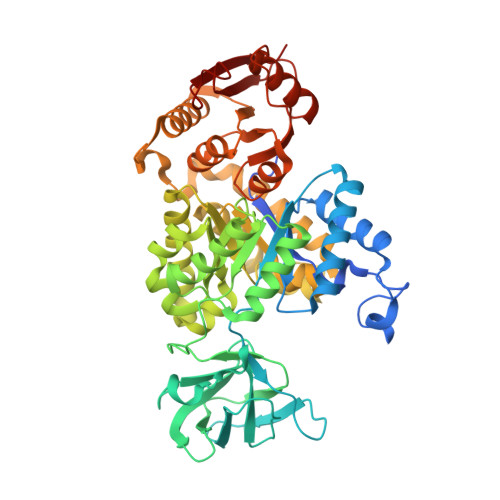
    Glycolysis is the primary metabolic pathway in the strictly fermentative Streptococcus pneumoniae, which is a major human pathogen associated with antibiotic resistance. Pyruvate kinase (PYK) is the last enzyme in this pathway that catalyzes the production of pyruvate from phosphoenolpyruvate (PEP) and plays a crucial role in controlling carbon flux; however, while S. pneumoniae PYK (SpPYK) is indispensable for growth, surprisingly little is known about its functional properties. Here, we report that compromising mutations in SpPYK confers resistance to the antibiotic fosfomycin, which inhibits the peptidoglycan synthesis enzyme MurA, implying a direct link between PYK and cell wall biogenesis. The crystal structures of SpPYK in the apo and ligand-bound states reveal key interactions that contribute to its conformational change as well as residues responsible for the recognition of PEP and the allosteric activator fructose 1,6-bisphosphate (FBP). Strikingly, FBP binding was observed at a location distinct from previously reported PYK effector binding sites. Furthermore, we show that SpPYK could be engineered to become more responsive to glucose 6-phosphate instead of FBP by sequence and structure-guided mutagenesis of the effector binding site. Together, our work sheds light on the regulatory mechanism of SpPYK and lays the groundwork for antibiotic development that targets this essential enzyme.


  • Organizational Affiliation

    SANKEN (The Institute of Scientific and Industrial Research), Osaka University, Ibaraki, Osaka, Japan; Graduate School of Pharmaceutical Sciences, Osaka University, Suita, Osaka, Japan. Electronic address: taguchi@sanken.osaka-u.ac.jp.


Macromolecules
Find similar proteins by:  (by identity cutoff)  |  3D Structure
Entity ID: 1
MoleculeChains Sequence LengthOrganismDetailsImage
Pyruvate kinase
A, B, C, D, E
A, B, C, D, E, F, G, H
521Streptococcus pneumoniae R6Mutation(s): 0 
Gene Names: pykF
UniProt
Find proteins for Q8DQ84 (Streptococcus pneumoniae (strain ATCC BAA-255 / R6))
Explore Q8DQ84 
Go to UniProtKB:  Q8DQ84
Entity Groups  
Sequence Clusters30% Identity50% Identity70% Identity90% Identity95% Identity100% Identity
UniProt GroupQ8DQ84
Sequence Annotations
Expand
  • Reference Sequence
Small Molecules
Ligands 7 Unique
IDChains Name / Formula / InChI Key2D Diagram3D Interactions
FBP
Query on FBP

Download Ideal Coordinates CCD File 
CA [auth E]
DA [auth F]
I [auth A]
IA [auth G]
N [auth B]
CA [auth E],
DA [auth F],
I [auth A],
IA [auth G],
N [auth B],
NA [auth H],
S [auth C],
X [auth D]
1,6-di-O-phosphono-beta-D-fructofuranose
C6 H14 O12 P2
RNBGYGVWRKECFJ-ARQDHWQXSA-N
PG4
Query on PG4

Download Ideal Coordinates CCD File 
HA [auth F]TETRAETHYLENE GLYCOL
C8 H18 O5
UWHCKJMYHZGTIT-UHFFFAOYSA-N
PGE
Query on PGE

Download Ideal Coordinates CCD File 
BA [auth D],
MA [auth G],
R [auth B],
W [auth C]
TRIETHYLENE GLYCOL
C6 H14 O4
ZIBGPFATKBEMQZ-UHFFFAOYSA-N
PEG
Query on PEG

Download Ideal Coordinates CCD File 
M [auth A],
RA [auth H]
DI(HYDROXYETHYL)ETHER
C4 H10 O3
MTHSVFCYNBDYFN-UHFFFAOYSA-N
OXL
Query on OXL

Download Ideal Coordinates CCD File 
FA [auth F]
K [auth A]
KA [auth G]
P [auth B]
PA [auth H]
FA [auth F],
K [auth A],
KA [auth G],
P [auth B],
PA [auth H],
U [auth C],
Z [auth D]
OXALATE ION
C2 O4
MUBZPKHOEPUJKR-UHFFFAOYSA-L
K
Query on K

Download Ideal Coordinates CCD File 
AA [auth D]
GA [auth F]
L [auth A]
LA [auth G]
Q [auth B]
AA [auth D],
GA [auth F],
L [auth A],
LA [auth G],
Q [auth B],
QA [auth H],
V [auth C]
POTASSIUM ION
K
NPYPAHLBTDXSSS-UHFFFAOYSA-N
MG
Query on MG

Download Ideal Coordinates CCD File 
EA [auth F]
J [auth A]
JA [auth G]
O [auth B]
OA [auth H]
EA [auth F],
J [auth A],
JA [auth G],
O [auth B],
OA [auth H],
T [auth C],
Y [auth D]
MAGNESIUM ION
Mg
JLVVSXFLKOJNIY-UHFFFAOYSA-N
Experimental Data & Validation

Experimental Data

  • Method: X-RAY DIFFRACTION
  • Resolution: 2.00 Å
  • R-Value Free: 0.246 
  • R-Value Work: 0.200 
  • R-Value Observed: 0.202 
  • Space Group: P 1
Unit Cell:
Length ( Å )Angle ( ˚ )
a = 73.442α = 88.95
b = 114.66β = 88.12
c = 140.705γ = 76.52
Software Package:
Software NamePurpose
REFMACrefinement
XDSdata scaling
XDSdata reduction
MOLREPphasing

Structure Validation

View Full Validation Report


Ligand Structure Quality Assessment 


Entry History & Funding Information

Deposition Data


Funding OrganizationLocationGrant Number
Japan Society for the Promotion of Science (JSPS)Japan21K20759

Revision History  (Full details and data files)

  • Version 1.0: 2023-06-14
    Type: Initial release
  • Version 1.1: 2023-06-21
    Changes: Database references
  • Version 1.2: 2023-07-19
    Changes: Data collection, Database references