Structural and interaction analysis of human lipocalin-type prostaglandin D synthase with the poorly water-soluble drug NBQX.
Miyamoto, Y., Nakatsuji, M., Yoshida, T., Ohkubo, T., Inui, T.(2023) FEBS J 290: 3983-3996
- PubMed: 37021622 Search on PubMed
- DOI: https://doi.org/10.1111/febs.16791
- Primary Citation Related Structures:
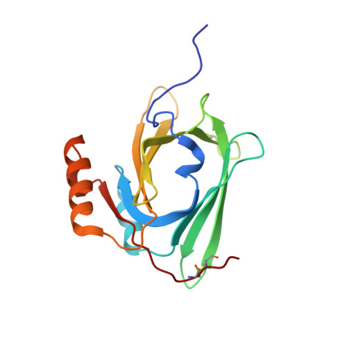
8HTA - PubMed Abstract:
Lipocalin-type prostaglandin D synthase (L-PGDS) is a secretory lipid-transporter protein that was shown to bind a wide variety of hydrophobic ligands in vitro. Exploiting this function, we previously examined the feasibility of using L-PGDS as a novel delivery vehicle for poorly water-soluble drugs. However, the mechanism by which human L-PGDS binds to poorly water-soluble drugs is unclear. In this study, we determined the solution structure of human L-PGDS and investigated the mechanism of L-PGDS binding to 6-nitro-7-sulfamoyl-benzo[f]quinoxalin-2,3-dione (NBQX), an α-amino-3-hydroxy-5-methyl-4-isoxazole-propionic acid receptor antagonist. NMR experiments showed that human L-PGDS has an eight-stranded antiparallel β-barrel structure that forms a central cavity, a short 3 10 -helix and two α-helices. Titration with NBQX was monitored using 1 H- 15 N HSQC spectroscopy. At higher NBQX concentrations, some cross-peaks of the protein exhibited fast-exchanging shifts with a curvature, indicating at least two binding sites. These residues were located in the upper portion of the cavity. Singular value decomposition analysis revealed that human L-PGDS has two NBQX binding sites. Large chemical shift changes were observed in the H2-helix and A-, B-, C-, D-, H- and I-strands and H2-helix upon NBQX binding. Calorimetric experiments revealed that human L-PGDS binds two NBQX molecules with dissociation constants of 46.7 μm for primary binding and 185.0 μm for secondary binding. Molecular docking simulations indicated that these NBQX binding sites are located within the β-barrel. These results provide new insights into the interaction between poorly water-soluble drugs and human L-PGDS as a drug carrier.
- Laboratory of Biological Macromolecules, Graduate School of Life and Environmental Sciences, Osaka Prefecture University, Sakai, Japan.
Organizational Affiliation: