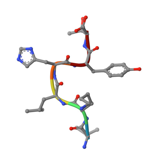
8HQT | pdb_00008hqt

The complex structure of COPI cargo sorting module with SARS-CoV-2 Spike KxHxx sorting motif


Experimental Data Snapshot

  • Method: X-RAY DIFFRACTION
  • Resolution: 1.80 Å
  • R-Value Free: 
    0.225 (Depositor), 0.226 (DCC) 
  • R-Value Work: 
    0.209 (Depositor), 0.210 (DCC) 
  • R-Value Observed: 
    0.210 (Depositor) 

Starting Model: experimental
View more details

wwPDB Validation 3D Report Full Report

Validation slider image for 8HQT

This is version 1.1 of the entry. See complete history. 

Literature

SARS-CoV-2 spike host cell surface exposure promoted by a COPI sorting inhibitor.

Li, Y., Yang, M., Nan, Y., Wang, J., Wang, S., Cui, D., Guo, J., He, P., Dai, W., Zhou, S., Zhang, Y., Ma, W.

(2023) Acta Pharm Sin B 13: 3043-3053

  • DOI: https://doi.org/10.1016/j.apsb.2023.04.007
  • Primary Citation Related Structures: 
    8HQT, 8HQV, 8HQW, 8HQX, 8HR0

  • PubMed Abstract: 

    Via an insufficient coat protein complex I (COPI) retrieval signal, the majority of SARS-CoV-2 spike (S) is resident in host early secretory organelles and a tiny amount is leaked out in cell surface. Only surface-exposed S can be recognized by B cell receptor (BCR) or anti-S therapeutic monoclonal antibodies (mAbs) that is the trigger step for B cell activation after S mRNA vaccination or infected cell clearance by S mAbs. Now, a drug strategy to promote S host surface exposure is absent. Here, we first combined structural and biochemical analysis to characterize S COPI sorting signals. A potent S COPI sorting inhibitor was then invented, evidently capable of promoting S surface exposure and facilitating infected cell clearance by S antibody-dependent cellular cytotoxicity (ADCC). Importantly, with the inhibitor as a probe, we revealed Omicron BA.1 S is less cell surface exposed than prototypes because of a constellation of S folding mutations, possibly corresponding to its ER chaperone association. Our findings not only suggest COPI is a druggable target against COVID-19, but also highlight SARS-CoV-2 evolution mechanism driven by S folding and trafficking mutations.


  • Organizational Affiliation: 
    • School of Life Science, Beijing University of Chinese Medicine, Beijing 102488, China.

Macromolecule Content 

  • Total Structure Weight: 35.06 kDa 
  • Atom Count: 2,605 
  • Modeled Residue Count: 301 
  • Deposited Residue Count: 307 
  • Unique protein chains: 2

Macromolecules

Find similar proteins by:|  3D Structure
Entity ID: 1
MoleculeChains  Sequence LengthOrganismDetailsImage
Coatomer subunit beta'301Saccharomyces cerevisiae YJM789Mutation(s): 0 
Gene Names: SEC27, SCY_1929
UniProt
Find proteins for P41811 (Saccharomyces cerevisiae (strain ATCC 204508 / S288c))
Explore P41811 
Go to UniProtKB:  P41811
Entity Groups
Sequence Clusters30% Identity50% Identity70% Identity90% Identity95% Identity100% Identity
UniProt GroupP41811
Sequence Annotations
Expand
Reference Sequence
Find similar proteins by:  Sequence   |   3D Structure  
Entity ID: 2
MoleculeChains  Sequence LengthOrganismDetailsImage
SARS-CoV-2 Spike KxHxx motif6Severe acute respiratory syndrome coronavirus 2Mutation(s): 0 
UniProt
Find proteins for P0DTC2 (Severe acute respiratory syndrome coronavirus 2)
Explore P0DTC2 
Go to UniProtKB:  P0DTC2
Entity Groups
UniProt GroupP0DTC2
Sequence Annotations
Expand
Reference Sequence

Experimental Data & Validation

Experimental Data

  • Method: X-RAY DIFFRACTION
  • Resolution: 1.80 Å
  • R-Value Free:  0.225 (Depositor), 0.226 (DCC) 
  • R-Value Work:  0.209 (Depositor), 0.210 (DCC) 
  • R-Value Observed: 0.210 (Depositor) 
Space Group: C 1 2 1
Unit Cell:
Length ( Å )Angle ( ˚ )
a = 74.892α = 90
b = 51.051β = 101.62
c = 85.101γ = 90
Software Package:
Software NamePurpose
PHENIXrefinement
HKL-3000data reduction
SCALAdata scaling
PHASERphasing

Structure Validation

View Full Validation Report


Entry History 

& Funding Information

Deposition Data


Funding OrganizationLocationGrant Number
Beijing University of Chinese Medicine (BUCM)China90011451310011, 1000061223476, ZYYCXTD-C-202006

Revision History  (Full details and data files)

  • Version 1.0: 2023-12-20
    Type: Initial release
  • Version 1.1: 2025-01-29
    Changes: Database references, Structure summary