Functional characterization, structural basis, and protein engineering of a rare flavonoid 2'- O -glycosyltransferase from Scutellaria baicalensis .
Wang, Z., Du, X., Ye, G., Wang, H., Liu, Y., Liu, C., Li, F., Agren, H., Zhou, Y., Li, J., He, C., Guo, D.A., Ye, M.(2024) Acta Pharm Sin B 14: 3746-3759
- PubMed: 39220864 Search on PubMedSearch on PubMed Central
- DOI: https://doi.org/10.1016/j.apsb.2024.04.001
- Primary Citation Related Structures:
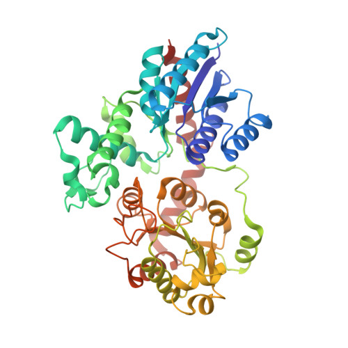
8HOJ, 8HOK - PubMed Abstract:
Glycosylation is an important post-modification reaction in plant secondary metabolism, and contributes to structural diversity of bioactive natural products. In plants, glycosylation is usually catalyzed by UDP-glycosyltransferases. Flavonoid 2'- O -glycosides are rare glycosides. However, no UGTs have been reported, thus far, to specifically catalyze 2'- O -glycosylation of flavonoids. In this work, UGT71AP2 was identified from the medicinal plant Scutellaria baicalensis as the first flavonoid 2'- O -glycosyltransferase. It could preferentially transfer a glycosyl moiety to 2'-hydroxy of at least nine flavonoids to yield six new compounds. Some of the 2'- O -glycosides showed noticeable inhibitory activities against cyclooxygenase 2. The crystal structure of UGT71AP2 (2.15 Å) was solved, and mechanisms of its regio-selectivity was interpreted by p K a calculations, molecular docking, MD simulation, MM/GBSA binding free energy, QM/MM, and hydrogen‒deuterium exchange mass spectrometry analysis. Through structure-guided rational design, we obtained the L138T/V179D/M180T mutant with remarkably enhanced regio-selectivity (the ratio of 7- O -glycosylation byproducts decreased from 48% to 4%) and catalytic efficiency of 2'- O -glycosylation ( k cat / K m , 0.23 L/(s·μmol), 12-fold higher than the native). Moreover, UGT71AP2 also possesses moderate UDP-dependent de-glycosylation activity, and is a dual function glycosyltransferase. This work provides an efficient biocatalyst and sets a good example for protein engineering to optimize enzyme catalytic features through rational design.
- State Key Laboratory of Natural and Biomimetic Drugs, School of Pharmaceutical Sciences, Peking University, Beijing 100191, China.
Organizational Affiliation: