Structural insights into the distinct substrate preferences of two bacterial epoxide hydrolases.
Hwang, J., Lee, M.J., Lee, S.G., Do, H., Lee, J.H.(2024) Int J Biol Macromol 264: 130419-130419
- PubMed: 38423431 Search on PubMed
- DOI: https://doi.org/10.1016/j.ijbiomac.2024.130419
- Primary Citation Related Structures:
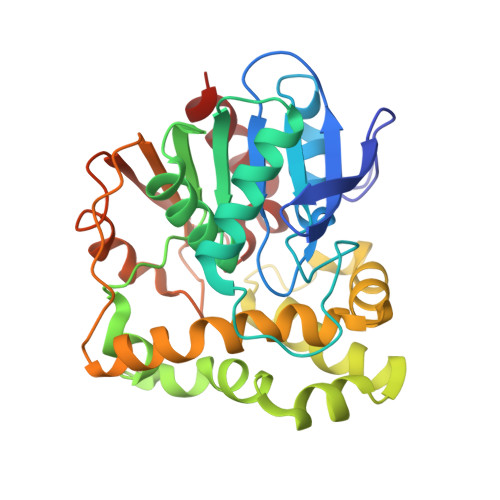
8HGU, 8HM5 - PubMed Abstract:
Epoxide hydrolases (EHs), which catalyze the transformation of epoxides to diols, are present in many eukaryotic and prokaryotic organisms. They have recently drawn considerable attention from organic chemists owing to their application in the semisynthesis of enantiospecific diol compounds. Here, we report the crystal structures of BoEH from Bosea sp. PAMC 26642 and CaEH from Caballeronia sordidicola PAMC 26510 at 1.95 and 2.43 Å resolution, respectively. Structural analysis showed that the overall structures of BoEH and CaEH commonly possess typical α/β hydrolase fold with the same ring-opening residues (Tyr-Tyr) and conserved catalytic triad residues (Asp-Asp-His). However, the two enzymes were found to have significantly different sequence compositions in the cap domain region, which is involved in the formation of the substrate-binding site in both enzymes. Enzyme activity assay results showed that BoEH had the strongest activity toward the linear aliphatic substrates, whereas CaEH had a higher preference for aromatic- and cycloaliphatic substrates. Computational docking simulations and tunnel identification revealed important residues with different substrate-binding preferences. Collectively, structure comparison studies, together with ligand docking simulation results, suggested that the differences in substrate-binding site residues were highly correlated with substrate specificity.
- Division of Life Sciences, Korea Polar Research Institute, Incheon 21990, Republic of Korea; Department of Polar Sciences, University of Science and Technology, Incheon 21990, Republic of Korea.
Organizational Affiliation: