Biochemical and Structural Analyses Shed Light on the Mechanisms of RadD DNA Binding and Its ATPase from Escherichia coli.
Tian, L.F., Kuang, X., Ding, K., Gao, H., Tang, Q., Yan, X.X., Xu, W.(2023) Int J Mol Sci 24
- PubMed: 36614183 Search on PubMedSearch on PubMed Central
- DOI: https://doi.org/10.3390/ijms24010741
- Primary Citation Related Structures:
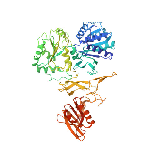
8H5Y, 8H5Z - PubMed Abstract:
DNA double-strand breaks (DSBs) are the most perilous and harmful type of DNA damage and can cause tumorigenesis or cell death if left repaired with an error or unrepaired. RadD, a member of the SF2 family, is a recently discovered DNA repair protein involved in the repair of DSBs after radiation or chemical damage. However, the function of RadD in DNA repair remains unclear. Here, we determined the crystal structures of RadD/ATPγS and RadD/ATP complexes and revealed the novel mechanism of RadD binding to DNA and ATP hydrolysis with biochemical data. In the RadD catalytic center, the Gly34 and Gly36 on the P-loop are key residues for ATP binding besides the conserved amino acids Lys37 and Arg343 in the SF2 family. If any of them mutate, then RadD loses ATPase activity. Asp117 polarizes the attacking water molecule, which then starts a nucleophilic reaction toward γ-phosphate, forming the transition state. Lys68 acts as a pocket switch to regulate substrate entry and product release. We revealed that the C-terminal peptide of single-stranded DNA-binding protein (SSB) binds the RadD C-terminal domain (CTD) and promotes the RadD ATPase activity. Our mutagenesis studies confirmed that the residues Arg428 on the zinc finger domain (ZFD) and Lys488 on the CTD of RadD are the key sites for binding branched DNA. Using the Coot software combined with molecular docking, we propose a RadD-binding DNA model for the DNA damage repair process.
- National Laboratory of Biomacromolecules, CAS Center for Excellence in Biomacromolecules, Institute of Biophysics, Chinese Academy of Sciences, Beijing 100101, China.
Organizational Affiliation: