8H1L

Crystal structure of glucose-2-epimerase in complex with D-Glucitol from Runella slithyformis Runsl_4512


Experimental Data Snapshot

  • Method: X-RAY DIFFRACTION
  • Resolution: 2.33 Å
  • R-Value Free: 0.208 
  • R-Value Work: 0.179 
  • R-Value Observed: 0.181 

wwPDB Validation   3D Report Full Report


Ligand Structure Quality Assessment 


This is version 1.1 of the entry. See complete history


Literature

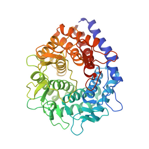
Structural insights into the substrate specificity and activity of a novel mannose 2-epimerase from Runella slithyformis.

Wang, H.Sun, X.Saburi, W.Hashiguchi, S.Yu, J.Ose, T.Mori, H.Yao, M.

(2023) Acta Crystallogr D Struct Biol 79: 585-595

  • DOI: https://doi.org/10.1107/S205979832300390X
  • Primary Citation of Related Structures:  
    8H1K, 8H1L, 8H1M, 8H1N

  • PubMed Abstract: 

    Mannose 2-epimerase (ME), a member of the acylglucosamine 2-epimerase (AGE) superfamily that catalyzes epimerization of D-mannose and D-glucose, has recently been characterized to have potential for D-mannose production. However, the substrate-recognition and catalytic mechanism of ME remains unknown. In this study, structures of Runella slithyformis ME (RsME) and its D254A mutant [RsME(D254A)] were determined in their apo forms and as intermediate-analog complexes [RsME-D-glucitol and RsME(D254A)-D-glucitol]. RsME possesses the (α/α) 6 -barrel of the AGE superfamily members but has a unique pocket-covering long loop (loop α7-α8 ). The RsME-D-glucitol structure showed that loop α7-α8 moves towards D-glucitol and closes the active pocket. Trp251 and Asp254 in loop α7-α8 are only conserved in MEs and interact with D-glucitol. Kinetic analyses of the mutants confirmed the importance of these residues for RsME activity. Moreover, the structures of RsME(D254A) and RsME(D254A)-D-glucitol revealed that Asp254 is vital for binding the ligand in a correct conformation and for active-pocket closure. Docking calculations and structural comparison with other 2-epimerases show that the longer loop α7-α8 in RsME causes steric hindrance upon binding to disaccharides. A detailed substrate-recognition and catalytic mechanism for monosaccharide-specific epimerization in RsME has been proposed.


  • Organizational Affiliation

    Faculty of Advanced Life Science, Hokkaido University, Kita 10, Nishi 8, Kita-ku, Sapporo 060-0810, Japan.


Macromolecules
Find similar proteins by:  (by identity cutoff)  |  3D Structure
Entity ID: 1
MoleculeChains Sequence LengthOrganismDetailsImage
N-acylglucosamine 2-epimerase
A, B, C, D
423Runella slithyformisMutation(s): 0 
Gene Names: Runsl_4512
UniProt
Find proteins for A0A7U4E834 (Runella slithyformis (strain ATCC 29530 / DSM 19594 / LMG 11500 / NCIMB 11436 / LSU 4))
Explore A0A7U4E834 
Go to UniProtKB:  A0A7U4E834
Entity Groups  
Sequence Clusters30% Identity50% Identity70% Identity90% Identity95% Identity100% Identity
UniProt GroupA0A7U4E834
Sequence Annotations
Expand
  • Reference Sequence
Experimental Data & Validation

Experimental Data

  • Method: X-RAY DIFFRACTION
  • Resolution: 2.33 Å
  • R-Value Free: 0.208 
  • R-Value Work: 0.179 
  • R-Value Observed: 0.181 
  • Space Group: C 1 2 1
Unit Cell:
Length ( Å )Angle ( ˚ )
a = 146.242α = 90
b = 172.731β = 116.964
c = 150.457γ = 90
Software Package:
Software NamePurpose
PHENIXrefinement
XDSdata reduction
XDSdata scaling
PHENIXphasing

Structure Validation

View Full Validation Report


Ligand Structure Quality Assessment 


Entry History & Funding Information

Deposition Data


Funding OrganizationLocationGrant Number
Japan Society for the Promotion of Science (JSPS)Japan18K05382
Japan Society for the Promotion of Science (JSPS)Japan21K05388
Japan Society for the Promotion of Science (JSPS)Japan21H01754
Japan Agency for Medical Research and Development (AMED)JapanJP18am0101071 (0058)
Japan Agency for Medical Research and Development (AMED)JapanJP19am0101083

Revision History  (Full details and data files)

  • Version 1.0: 2023-07-12
    Type: Initial release
  • Version 1.1: 2023-11-29
    Changes: Data collection, Refinement description