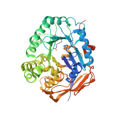
Complexed Crystal Structure of the Dihydroorotase Domain of Human CAD Protein with the Anticancer Drug 5-Fluorouracil.
Lin, E.S., Huang, Y.H., Yang, P.C., Peng, W.F., Huang, C.Y.(2023) Biomolecules 13
- PubMed: 36671534 Search on PubMedSearch on PubMed Central
- DOI: https://doi.org/10.3390/biom13010149
- Primary Citation Related Structures:
8GVZ, 8GW0 - PubMed Abstract:
Dihydroorotase (DHOase) is the third enzyme in the pathway used for the biosynthesis of pyrimidine nucleotides. In mammals, DHOase is active in a trifunctional enzyme, CAD, which also carries out the activities of carbamoyl phosphate synthetase and aspartate transcarbamoylase. Prior to this study, it was unknown whether the FDA-approved clinical drug 5-fluorouracil (5-FU), which is used as an anticancer therapy, could bind to the DHOase domain of human CAD (huDHOase). Here, we identified huDHOase as a new 5-FU binding protein, thereby extending the 5-FU interactome to this human enzyme. In order to investigate where 5-FU binds to huDHOase, we solved the complexed crystal structure at 1.97 Å (PDB ID 8GVZ). The structure of huDHOase complexed with malate was also determined for the sake of comparison (PDB ID 8GW0). These two nonsubstrate ligands were bound at the active site of huDHOase. It was previously established that the substrate N -carbamoyl-L-aspartate is either bound to or moves away from the active site, but it is the loop that is extended towards (loop-in mode) or moved away (loop-out mode) from the active site. DHOase also binds to nonsubstrate ligands via the loop-out mode. In contrast to the Escherichia coli DHOase model, our complexed structures revealed that huDHOase binds to either 5-FU or malate via the loop-in mode. We further characterized the binding of 5-FU to huDHOase using site-directed mutagenesis and the fluorescence quenching method. Considering the loop-in mode, the dynamic loop in huDHOase should be a suitable drug-targeting site for further designing inhibitors and clinical chemotherapies to suppress pyrimidine biosynthesis in cancer cell lines.
- Department of Beauty Science, National Taichung University of Science and Technology, Taichung City 403, Taiwan.
Organizational Affiliation: