8GTL

Crystal Structure of Cytochrome P450 (CYP101D5)


Experimental Data Snapshot

  • Method: X-RAY DIFFRACTION
  • Resolution: 3.20 Å
  • R-Value Free: 0.319 
  • R-Value Work: 0.262 
  • R-Value Observed: 0.268 

wwPDB Validation   3D Report Full Report


Ligand Structure Quality Assessment 


This is version 1.1 of the entry. See complete history


Literature

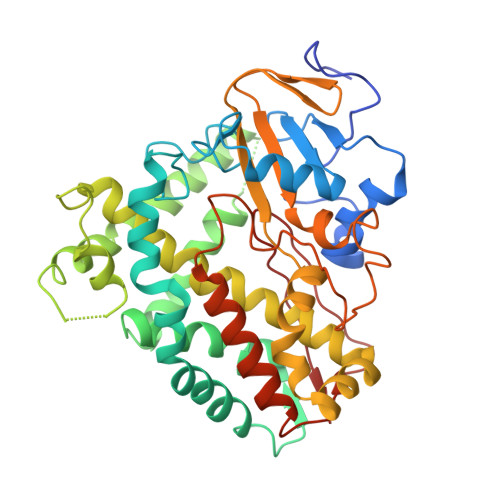
Crystal Structure and Biochemical Analysis of a Cytochrome P450 CYP101D5 from Sphingomonas echinoides.

Subedi, P.Do, H.Lee, J.H.Oh, T.J.

(2022) Int J Mol Sci 23

  • DOI: https://doi.org/10.3390/ijms232113317
  • Primary Citation of Related Structures:  
    8GTL

  • PubMed Abstract: 

    Cytochrome P450 enzymes (CYPs) are heme-containing enzymes that catalyze hydroxylation with a variety of biological molecules. Despite their diverse activity and substrates, the structures of CYPs are limited to a tertiary structure that is similar across all the enzymes. It has been presumed that CYPs overcome substrate selectivity with highly flexible loops and divergent sequences around the substrate entrance region. Here, we report the newly identified CYP101D5 from Sphingomonas echinoides . CYP101D5 catalyzes the hydroxylation of β-ionone and flavonoids, including naringenin and apigenin, and causes the dehydrogenation of α-ionone. A structural investigation and comparison with other CYP101 families indicated that spatial constraints at the substrate-recognition site originate from the B/C loop. Furthermore, charge distribution at the substrate binding site may be important for substrate selectivity and the preference for CYP101D5.


  • Organizational Affiliation

    Department of Life Science and Biochemical Engineering, Graduate School, Sun Moon University, Asan 31460, Korea.


Macromolecules
Find similar proteins by:  (by identity cutoff)  |  3D Structure
Entity ID: 1
MoleculeChains Sequence LengthOrganismDetailsImage
cytochrome P450 CYP101D5
A, B
409Sphingomonas echinoidesMutation(s): 0 
Entity Groups  
Sequence Clusters30% Identity50% Identity70% Identity90% Identity95% Identity100% Identity
Sequence Annotations
Expand
  • Reference Sequence
Small Molecules
Experimental Data & Validation

Experimental Data

  • Method: X-RAY DIFFRACTION
  • Resolution: 3.20 Å
  • R-Value Free: 0.319 
  • R-Value Work: 0.262 
  • R-Value Observed: 0.268 
  • Space Group: P 21 21 21
Unit Cell:
Length ( Å )Angle ( ˚ )
a = 68.524α = 90
b = 109.573β = 90
c = 113.878γ = 90
Software Package:
Software NamePurpose
Blu-Icedata collection
PHENIXrefinement
Blu-Icedata collection
HKL-2000data reduction
HKL-2000data scaling
MOLREPphasing

Structure Validation

View Full Validation Report


Ligand Structure Quality Assessment 


Entry History & Funding Information

Deposition Data


Funding OrganizationLocationGrant Number
Ministry of Education (MoE, Korea)Korea, Republic OfNRF-2019R1D1A3A03103903

Revision History  (Full details and data files)

  • Version 1.0: 2022-12-28
    Type: Initial release
  • Version 1.1: 2023-11-29
    Changes: Data collection, Refinement description