8GHO

GUCY2C-peptide bound to anti-GUCY2C-scFv antibody


Experimental Data Snapshot

  • Method: X-RAY DIFFRACTION
  • Resolution: 1.60 Å
  • R-Value Free: 0.217 
  • R-Value Work: 0.197 
  • R-Value Observed: 0.198 

wwPDB Validation   3D Report Full Report


This is version 1.1 of the entry. See complete history


Literature

Molecular insights into recognition of GUCY2C by T-cell engaging bispecific antibody anti-GUCY2CxCD3.

Rampuria, P.Mosyak, L.Root, A.R.Svenson, K.Agostino, M.J.LaVallie, E.R.

(2023) Sci Rep 13: 13408-13408

  • DOI: https://doi.org/10.1038/s41598-023-40467-0
  • Primary Citation of Related Structures:  
    8GHO, 8GHP

  • PubMed Abstract: 

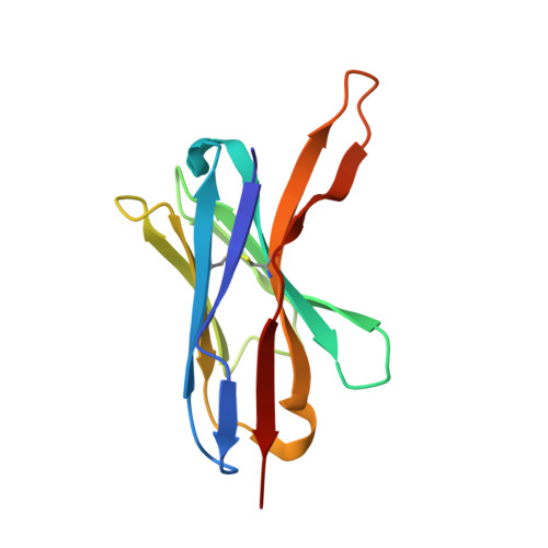
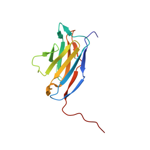
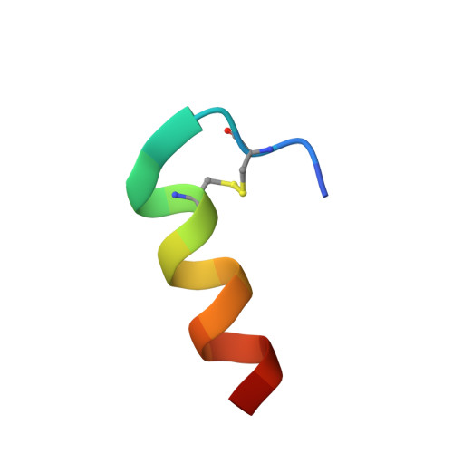
    The intestinal epithelial receptor Guanylyl Cyclase C (GUCY2C) is a tumor-associated cell surface antigen expressed across gastrointestinal malignancies that can serve as an efficacious target for colorectal cancer immunotherapy. Here, we describe a yeast surface-display approach combined with an orthogonal peptide-based mapping strategy to identify the GUCY2C binding epitope of a novel anti-GUCY2CxCD3 bispecific antibody (BsAb) that recently advanced into the clinic for the treatment of cancer. The target epitope was localized to the N-terminal helix H2 of human GUCY2C, which enabled the determination of the crystal structure of the minimal GUCY2C epitope in complex with the anti-GUCY2C antibody domain. To understand if this minimal epitope covers the entire antibody binding region and to investigate the impact of epitope position on the antibody's activity, we further determined the structure of this interaction in the context of the full-length extracellular domain (ECD) of GUCY2C. We found that this epitope is positioned on the protruding membrane-distal helical region of GUCY2C and that its specific location on the surface of GUCY2C dictates the close spatial proximity of the two antigen arms in a diabody arrangement essential to the tumor killing activity of GUCY2CxCD3 BsAb.


  • Organizational Affiliation

    Biomedicine Design, Pfizer Inc., 610 Main St., Cambridge, MA, 02139, USA. pragya.rampuria@pfizer.com.


Macromolecules
Find similar proteins by:  (by identity cutoff)  |  3D Structure
Entity ID: 1
MoleculeChains Sequence LengthOrganismDetailsImage
anti-GUCY2C-scFv antibody heavy chainA,
E [auth H]
122Homo sapiensMutation(s): 0 
Entity Groups  
Sequence Clusters30% Identity50% Identity70% Identity90% Identity95% Identity100% Identity
Sequence Annotations
Expand
  • Reference Sequence
Find similar proteins by:  (by identity cutoff)  |  3D Structure
Entity ID: 2
MoleculeChains Sequence LengthOrganismDetailsImage
anti-GUCY2C-scFv antibody light chainB,
F [auth L]
121Homo sapiensMutation(s): 0 
Entity Groups  
Sequence Clusters30% Identity50% Identity70% Identity90% Identity95% Identity100% Identity
Sequence Annotations
Expand
  • Reference Sequence

Find similar proteins by:  Sequence   |   3D Structure  

Entity ID: 3
MoleculeChains Sequence LengthOrganismDetailsImage
Guanylyl cyclase C peptideC,
D [auth G]
18Homo sapiensMutation(s): 0 
EC: 4.6.1.2
UniProt & NIH Common Fund Data Resources
Find proteins for P25092 (Homo sapiens)
Explore P25092 
Go to UniProtKB:  P25092
PHAROS:  P25092
GTEx:  ENSG00000070019 
Entity Groups  
Sequence Clusters30% Identity50% Identity70% Identity90% Identity95% Identity100% Identity
UniProt GroupP25092
Sequence Annotations
Expand
  • Reference Sequence
Experimental Data & Validation

Experimental Data

  • Method: X-RAY DIFFRACTION
  • Resolution: 1.60 Å
  • R-Value Free: 0.217 
  • R-Value Work: 0.197 
  • R-Value Observed: 0.198 
  • Space Group: P 21 21 21
Unit Cell:
Length ( Å )Angle ( ˚ )
a = 70.68α = 90
b = 80.57β = 90
c = 90.7γ = 90
Software Package:
Software NamePurpose
STARANISOdata scaling
BUSTERrefinement
autoPROCdata reduction
PHASERphasing

Structure Validation

View Full Validation Report


Entry History & Funding Information

Deposition Data

  • Released Date: 2023-08-30 
  • Deposition Author(s): Mosyak, L.

Funding OrganizationLocationGrant Number
Not funded--

Revision History  (Full details and data files)

  • Version 1.0: 2023-08-30
    Type: Initial release
  • Version 1.1: 2023-10-11
    Changes: Refinement description