8GHH

Crystal structure of Staphylococcus aureus Lysophosphatidylglycerol phospholipase D

  • Classification: HYDROLASE
  • Organism(s): Staphylococcus aureus
  • Expression System: Escherichia coli
  • Mutation(s): No 

  • Deposited: 2023-03-10 Released: 2023-11-01 
  • Deposition Author(s): Rock, C.O., Yun, M.
  • Funding Organization(s): National Institutes of Health/National Institute of General Medical Sciences (NIH/NIGMS)

Experimental Data Snapshot

  • Method: X-RAY DIFFRACTION
  • Resolution: 2.10 Å
  • R-Value Free: 0.215 
  • R-Value Work: 0.188 
  • R-Value Observed: 0.189 

wwPDB Validation   3D Report Full Report


This is version 1.0 of the entry. See complete history


Literature

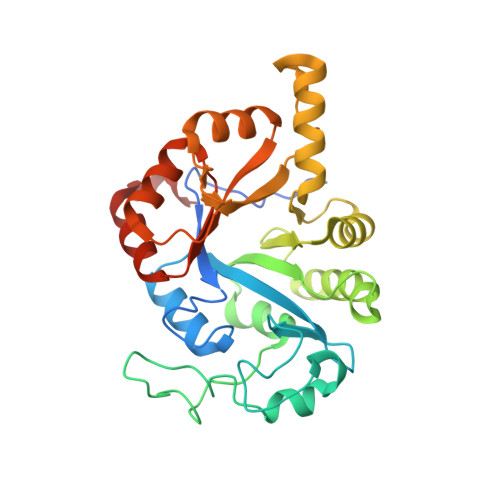
Lysophosphatidylglycerol (LPG) phospholipase D maintains membrane homeostasis in Staphylococcus aureus by converting LPG to lysophosphatidic acid.

Subramanian, C.Yun, M.K.Frank, M.M.Rock, C.O.

(2023) J Biol Chem 299: 104863-104863

  • DOI: https://doi.org/10.1016/j.jbc.2023.104863
  • Primary Citation of Related Structures:  
    8GHH, 8GHI

  • PubMed Abstract: 

    Lysophospholipids are deacylated derivatives of their bilayer forming phospholipid counterparts that are present at low concentrations in cells. Phosphatidylglycerol (PG) is the principal membrane phospholipid in Staphylococcus aureus and lysophosphatidylglycerol (LPG) is detected in low abundance. Here, we used a mass spectrometry screen to identify locus SAUSA300_1020 as the gene responsible for maintaining low concentrations of 1-acyl-LPG in S. aureus. The SAUSA300_1020 gene encodes a protein with a predicted amino terminal transmembrane α-helix attached to a globular glycerophosphodiester phosphodiesterase (GDPD) domain. We determined that the purified protein lacking the hydrophobic helix (LpgDΔN) possesses cation-dependent lysophosphatidylglycerol phospholipase D activity that generates both lysophosphatidic acid (LPA) and cyclic-LPA products and hydrolyzes cyclic-LPA to LPA. Mn 2+ was the highest affinity cation and stabilized LpgDΔN to thermal denaturation. LpgDΔN was not specific for the phospholipid headgroup and degraded 1-acyl-LPG, but not 2-acyl-LPG. Furthermore, a 2.1 Å crystal structure shows that LpgDΔN adopts the GDPD variation of the TIM barrel architecture except for the length and positioning of helix α6 and sheet β7. These alterations create a hydrophobic diffusion path for LPG to access the active site. The LpgD active site has the canonical GDPD metal binding and catalytic residues, and our biochemical characterization of site-directed mutants support a two-step mechanism involving a cyclic-LPA intermediate. Thus, the physiological function of LpgD in S. aureus is to convert LPG to LPA, which is re-cycled into the PG biosynthetic pathway at the LPA acyltransferase step to maintain membrane PG molecular species homeostasis.


  • Organizational Affiliation

    Department of Infectious Diseases, St Jude Children's Research Hospital, Memphis, Tennessee, USA.


Macromolecules
Find similar proteins by:  (by identity cutoff)  |  3D Structure
Entity ID: 1
MoleculeChains Sequence LengthOrganismDetailsImage
Glycerophosphodiester phosphodiesterase294Staphylococcus aureusMutation(s): 0 
Gene Names: 
EC: 3.1.4.46
UniProt
Find proteins for A0A0D6HT57 (Staphylococcus aureus)
Explore A0A0D6HT57 
Go to UniProtKB:  A0A0D6HT57
Entity Groups  
Sequence Clusters30% Identity50% Identity70% Identity90% Identity95% Identity100% Identity
UniProt GroupA0A0D6HT57
Sequence Annotations
Expand
  • Reference Sequence
Small Molecules
Ligands 3 Unique
IDChains Name / Formula / InChI Key2D Diagram3D Interactions
2HP
Query on 2HP

Download Ideal Coordinates CCD File 
B [auth A],
C [auth A]
DIHYDROGENPHOSPHATE ION
H2 O4 P
NBIIXXVUZAFLBC-UHFFFAOYSA-M
GOL
Query on GOL

Download Ideal Coordinates CCD File 
D [auth A]
E [auth A]
F [auth A]
G [auth A]
H [auth A]
D [auth A],
E [auth A],
F [auth A],
G [auth A],
H [auth A],
I [auth A],
J [auth A]
GLYCEROL
C3 H8 O3
PEDCQBHIVMGVHV-UHFFFAOYSA-N
CL
Query on CL

Download Ideal Coordinates CCD File 
K [auth A]CHLORIDE ION
Cl
VEXZGXHMUGYJMC-UHFFFAOYSA-M
Experimental Data & Validation

Experimental Data

  • Method: X-RAY DIFFRACTION
  • Resolution: 2.10 Å
  • R-Value Free: 0.215 
  • R-Value Work: 0.188 
  • R-Value Observed: 0.189 
  • Space Group: I 4 2 2
Unit Cell:
Length ( Å )Angle ( ˚ )
a = 107.956α = 90
b = 107.956β = 90
c = 138.381γ = 90
Software Package:
Software NamePurpose
PHENIXrefinement
HKL-2000data reduction
HKL-2000data scaling
PHASERphasing

Structure Validation

View Full Validation Report


Entry History & Funding Information

Deposition Data


Funding OrganizationLocationGrant Number
National Institutes of Health/National Institute of General Medical Sciences (NIH/NIGMS)United StatesGM034496

Revision History  (Full details and data files)

  • Version 1.0: 2023-11-01
    Type: Initial release