The structure of NAD + consuming protein Acinetobacter baumannii TIR domain shows unique kinetics and conformations.
Klontz, E., Obi, J.O., Wang, Y., Glendening, G., Carr, J., Tsibouris, C., Buddula, S., Nallar, S., Soares, A.S., Beckett, D., Redzic, J.S., Eisenmesser, E., Palm, C., Schmidt, K., Scudder, A.H., Obiorah, T., Essuman, K., Milbrandt, J., Diantonio, A., Ray, K., Snyder, M.L.D., Deredge, D., Snyder, G.A.(2023) J Biol Chem 299: 105290-105290
- PubMed: 37758001 Search on PubMedSearch on PubMed Central
- DOI: https://doi.org/10.1016/j.jbc.2023.105290
- Primary Citation Related Structures:
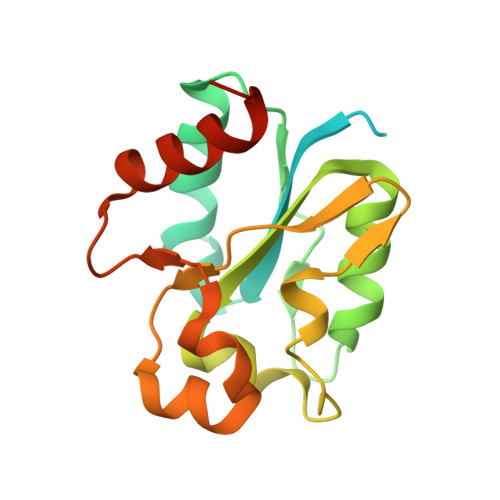
8G83 - PubMed Abstract:
Toll-like and interleukin-1/18 receptor/resistance (TIR) domain-containing proteins function as important signaling and immune regulatory molecules. TIR domain-containing proteins identified in eukaryotic and prokaryotic species also exhibit NAD+ hydrolase activity in select bacteria, plants, and mammalian cells. We report the crystal structure of the Acinetobacter baumannii TIR domain protein (AbTir-TIR) with confirmed NAD + hydrolysis and map the conformational effects of its interaction with NAD + using hydrogen-deuterium exchange-mass spectrometry. NAD + results in mild decreases in deuterium uptake at the dimeric interface. In addition, AbTir-TIR exhibits EX1 kinetics indicative of large cooperative conformational changes, which are slowed down upon substrate binding. Additionally, we have developed label-free imaging using the minimally invasive spectroscopic method 2-photon excitation with fluorescence lifetime imaging, which shows differences in bacteria expressing native and mutant NAD+ hydrolase-inactivated AbTir-TIR E208A protein. Our observations are consistent with substrate-induced conformational changes reported in other TIR model systems with NAD+ hydrolase activity. These studies provide further insight into bacterial TIR protein mechanisms and their varying roles in biology.
- Division of Vaccine Research, Institute of Human Virology, School of Medicine, University of Maryland, Baltimore, Maryland, USA.
Organizational Affiliation: