Cofactorless oxygenases guide anthraquinone-fused enediyne biosynthesis.
Gui, C., Kalkreuter, E., Liu, Y.C., Li, G., Steele, A.D., Yang, D., Chang, C., Shen, B.(2024) Nat Chem Biol 20: 243-250
- PubMed: 37945897 Search on PubMedSearch on PubMed Central
- DOI: https://doi.org/10.1038/s41589-023-01476-2
- Primary Citation Related Structures:
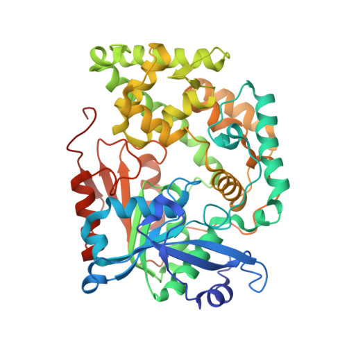
8G5S, 8G5T, 8G5U - PubMed Abstract:
The anthraquinone-fused enediynes (AFEs) combine an anthraquinone moiety and a ten-membered enediyne core capable of generating a cytotoxic diradical species. AFE cyclization is triggered by opening the F-ring epoxide, which is also the site of the most structural diversity. Previous studies of tiancimycin A, a heavily modified AFE, have revealed a cryptic aldehyde blocking installation of the epoxide, and no unassigned oxidases could be predicted within the tnm biosynthetic gene cluster. Here we identify two consecutively acting cofactorless oxygenases derived from methyltransferase and α/β-hydrolase protein folds, TnmJ and TnmK2, respectively, that are responsible for F-ring tailoring in tiancimycin biosynthesis by comparative genomics. Further biochemical and structural characterizations reveal that the electron-rich AFE anthraquinone moiety assists in catalyzing deformylation, epoxidation and oxidative ring cleavage without exogenous cofactors. These enzymes therefore fill important knowledge gaps for the biosynthesis of this class of molecules and the underappreciated family of cofactorless oxygenases.
- Department of Chemistry, The Herbert Wertheim UF Scripps Institute for Biomedical Innovation & Technology, University of Florida, Jupiter, FL, USA.
Organizational Affiliation: