8FG6

Design of amyloidogenic peptide traps


Experimental Data Snapshot

  • Method: X-RAY DIFFRACTION
  • Resolution: 2.30 Å
  • R-Value Free: 0.260 
  • R-Value Work: 0.212 
  • R-Value Observed: 0.215 

wwPDB Validation   3D Report Full Report


This is version 1.1 of the entry. See complete history


Literature

Design of amyloidogenic peptide traps.

Sahtoe, D.D.Andrzejewska, E.A.Han, H.L.Rennella, E.Schneider, M.M.Meisl, G.Ahlrichs, M.Decarreau, J.Nguyen, H.Kang, A.Levine, P.Lamb, M.Li, X.Bera, A.K.Kay, L.E.Knowles, T.P.J.Baker, D.

(2024) Nat Chem Biol 

  • DOI: https://doi.org/10.1038/s41589-024-01578-5
  • Primary Citation of Related Structures:  
    8FG6

  • PubMed Abstract: 

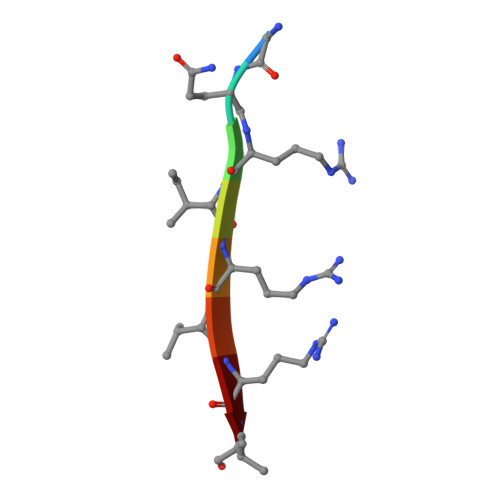
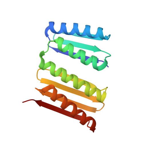
    Segments of proteins with high β-strand propensity can self-associate to form amyloid fibrils implicated in many diseases. We describe a general approach to bind such segments in β-strand and β-hairpin conformations using de novo designed scaffolds that contain deep peptide-binding clefts. The designs bind their cognate peptides in vitro with nanomolar affinities. The crystal structure of a designed protein-peptide complex is close to the design model, and NMR characterization reveals how the peptide-binding cleft is protected in the apo state. We use the approach to design binders to the amyloid-forming proteins transthyretin, tau, serum amyloid A1 and amyloid β 1-42 (Aβ42). The Aβ binders block the assembly of Aβ fibrils as effectively as the most potent of the clinically tested antibodies to date and protect cells from toxic Aβ42 species.


  • Organizational Affiliation

    Department of Biochemistry, University of Washington, Seattle, WA, USA. d.sahtoe@hubrecht.eu.


Macromolecules

Find similar proteins by:  Sequence   |   3D Structure  

Entity ID: 1
MoleculeChains Sequence LengthOrganismDetailsImage
amyloidogenic peptide8synthetic constructMutation(s): 0 
Sequence Annotations
Expand
  • Reference Sequence
Find similar proteins by:  (by identity cutoff)  |  3D Structure
Entity ID: 2
MoleculeChains Sequence LengthOrganismDetailsImage
C104.1152synthetic constructMutation(s): 0 
Entity Groups  
Sequence Clusters30% Identity50% Identity70% Identity90% Identity95% Identity100% Identity
Sequence Annotations
Expand
  • Reference Sequence
Experimental Data & Validation

Experimental Data

  • Method: X-RAY DIFFRACTION
  • Resolution: 2.30 Å
  • R-Value Free: 0.260 
  • R-Value Work: 0.212 
  • R-Value Observed: 0.215 
  • Space Group: P 31 2 1
Unit Cell:
Length ( Å )Angle ( ˚ )
a = 64.54α = 90
b = 64.54β = 90
c = 112.618γ = 120
Software Package:
Software NamePurpose
REFMACrefinement
Aimlessdata scaling
PHASERphasing
XDSdata reduction

Structure Validation

View Full Validation Report


Entry History & Funding Information

Deposition Data


Funding OrganizationLocationGrant Number
Howard Hughes Medical Institute (HHMI)United States--

Revision History  (Full details and data files)

  • Version 1.0: 2024-03-20
    Type: Initial release
  • Version 1.1: 2024-04-03
    Changes: Database references