Structural and functional insights of the catalytic GH5 and Calx-beta domains from the metagenome-derived endoglucanase CelE2.
Pimentel, A.C., Liberato, M.V., Franco Cairo, J.P.L., Tomazetto, G., Gandin, C.A., de Oliveira Neto, M., Alvarez, T.M., Squina, F.M.(2023) Enzyme Microb Technol 165: 110206-110206
- PubMed: 36758494 Search on PubMed
- DOI: https://doi.org/10.1016/j.enzmictec.2023.110206
- Primary Citation Related Structures:
8FC0 - PubMed Abstract:
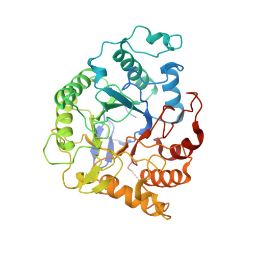
Cellulose is the most abundant natural polymer on Earth, representing an attractive feedstock for bioproducts and biofuel production. Cellulases promote the depolymerization of cellulose, generating short oligosaccharides and glucose, which are useful in biotechnological applications. Among the classical cellulases, those from glycoside hydrolase family 5 (GH5) are one of the most abundant in Nature, displaying several modular architectures with other accessory domains attached to its catalytic core, such as carbohydrate-binding modules (CBMs), Ig-like, FN3-like, and Calx-β domains, which can influence the enzyme activity. The metagenome-derived endoglucanase CelE2 has in its modular architecture an N-terminal domain belonging to the GH5 family and a C-terminal domain with a high identity to the Calx-β domain. In this study, the GH5 and the Calx-β domains were subcloned and heterologously expressed in E. coli, to evaluate the structural and functional properties of the individualized domains of CelE2. Thermostability analysis by circular dichroism (CD) revealed a decrease in the denaturation temperature values around 4.6 °C for the catalytic domain (CelE2 1-381 ) compared to CelE2 full-length. The CD analyses revealed that the Calx-β domain (CelE2 382-477 ) was unfolded, suggesting that this domain requires to be attached to the catalytic core to become structurally stable. The three-dimensional structure of the catalytic domain CelE2 1-381 was determined at 2.1 Å resolution, showing a typical (α/β) 8 -barrel fold and a narrow active site compared to other cellulases from the same family. The biochemical characterization showed that the deletion of the Calx-β domain increased more than 3-fold the activity of the catalytic domain CelE2 1-381 towards the insoluble substrate Avicel. The main functional properties of CelE2, such as substrate specificity, optimal pH and temperature, thermal stability, and activation by CaCl 2, were not altered after the deletion of the accessory domain. Furthermore, the Small Angle X-ray Scattering (SAXS) analyses showed that the addition of CaCl 2 was beneficial CelE2 1-381 protein solvency. This work contributed to fundamental concepts about the structure and function of cellulases, which are useful in applications involving lignocellulosic materials degradation into food and feedstuffs and biofuel production.
- Departamento de Bioquímica, Instituto de Biologia (IB), Universidade Estadual de Campinas (UNICAMP), R. Monteiro Lobato, 255- Cidade Universitária, Campinas, SP, Brasil. Electronic address: agnescristinap@gmail.com.
Organizational Affiliation: