8F27

Mirror-image DNA containing 2'-OMe-L-uridine residue


Experimental Data Snapshot

  • Method: X-RAY DIFFRACTION
  • Resolution: 1.60 Å
  • R-Value Free: 0.205 
  • R-Value Work: 0.193 
  • R-Value Observed: 0.193 

wwPDB Validation   3D Report Full Report


This is version 1.2 of the entry. See complete history


Literature

Derivatization of Mirror-Image l-Nucleic Acids with 2'-OMe Modification for Thermal and Structural Stabilization.

Dantsu, Y.Zhang, W.

(2023) Chembiochem 24: e202200764-e202200764

  • DOI: https://doi.org/10.1002/cbic.202200764
  • Primary Citation of Related Structures:  
    8F24, 8F27

  • PubMed Abstract: 

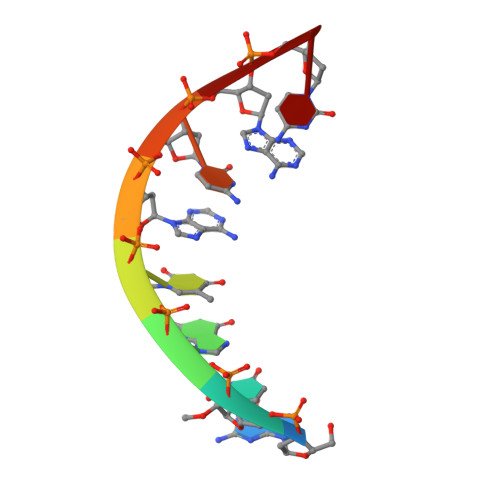
    To further expand the functionality and enhance the stability of mirror-image nucleic acids as advanced agents for basic research and therapeutic design, we have synthesized 2'-deoxy-2'-methoxy-l-uridine phosphoramidite and incorporated it into l-DNA and l-RNA by solid-phase synthesis quantitatively. We found that the thermostability of l-nucleic acids is dramatically improved after introducing the modifications. Moreover, we successfully crystallized both l-DNA and l-RNA duplexes containing the 2'-OMe modifications and sharing identical sequences. Crystal structure determination and analysis revealed the overall structures of the mirror-image nucleic acids, and for the first time it was possible to interpret the structural deviations caused by 2'-OMe and 2'-OH groups in the oligonucleotides, which are very similar. This novel chemical nucleic acid modification has the potential to be used to design nucleic acid-based therapeutics and materials in the future.


  • Organizational Affiliation

    Department of Biochemistry and Molecular Biology, Indiana University School of Medicine, 635 Barnhill Drive, Indianapolis, IN, 46202, USA.


Macromolecules

Find similar nucleic acids by:  Sequence   |   3D Structure  

Entity ID: 1
MoleculeChains LengthOrganismImage
Mirror-image DNA (5'-D(*(0DG)P*(0MU)P*(0DG)P*(0DT)P*(0DA)P*(0DC)P*(0DA)P*(0DC))-3')
A, B
8synthetic construct
Sequence Annotations
Expand
  • Reference Sequence
Small Molecules
Ligands 1 Unique
IDChains Name / Formula / InChI Key2D Diagram3D Interactions
SO4
Query on SO4

Download Ideal Coordinates CCD File 
C [auth B]SULFATE ION
O4 S
QAOWNCQODCNURD-UHFFFAOYSA-L
Experimental Data & Validation

Experimental Data

  • Method: X-RAY DIFFRACTION
  • Resolution: 1.60 Å
  • R-Value Free: 0.205 
  • R-Value Work: 0.193 
  • R-Value Observed: 0.193 
  • Space Group: P 65
Unit Cell:
Length ( Å )Angle ( ˚ )
a = 46.31α = 90
b = 46.31β = 90
c = 43.52γ = 120
Software Package:
Software NamePurpose
REFMACrefinement
HKL-2000data reduction
HKL-2000data scaling
PHASERphasing

Structure Validation

View Full Validation Report


Entry History & Funding Information

Deposition Data


Funding OrganizationLocationGrant Number
Not funded--

Revision History  (Full details and data files)

  • Version 1.0: 2023-04-12
    Type: Initial release
  • Version 1.1: 2023-06-28
    Changes: Database references
  • Version 1.2: 2023-10-25
    Changes: Data collection, Refinement description