8EYB

Crystal structure of PTP1B D181A/Q262A/C215A phosphatase domain with JAK2 activation loop phosphopeptide


Experimental Data Snapshot

  • Method: X-RAY DIFFRACTION
  • Resolution: 2.35 Å
  • R-Value Free: 0.243 
  • R-Value Work: 0.207 
  • R-Value Observed: 0.209 

wwPDB Validation   3D Report Full Report


Ligand Structure Quality Assessment 


This is version 1.2 of the entry. See complete history


Literature

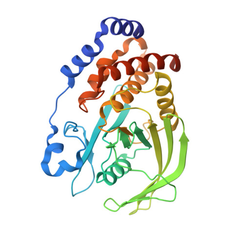
Structure guided studies of the interaction between PTP1B and JAK.

Morris, R.Keating, N.Tan, C.Chen, H.Laktyushin, A.Saiyed, T.Liau, N.P.D.Nicola, N.A.Tiganis, T.Kershaw, N.J.Babon, J.J.

(2023) Commun Biol 6: 641-641

  • DOI: https://doi.org/10.1038/s42003-023-05020-9
  • Primary Citation of Related Structures:  
    8EXI, 8EXJ, 8EXK, 8EXM, 8EXN, 8EYA, 8EYB, 8EYC, 8F88

  • PubMed Abstract: 

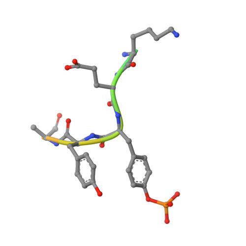
    Protein Tyrosine Phosphatase 1B (PTP1B) is the prototypical protein tyrosine phosphatase and plays an essential role in the regulation of several kinase-driven signalling pathways. PTP1B displays a preference for bisphosphorylated substrates. Here we identify PTP1B as an inhibitor of IL-6 and show that, in vitro, it can dephosphorylate all four members of the JAK family. In order to gain a detailed understanding of the molecular mechanism of JAK dephosphorylation, we undertook a structural and biochemical analysis of the dephosphorylation reaction. We identified a product-trapping PTP1B mutant that allowed visualisation of the tyrosine and phosphate products of the reaction and a substrate-trapping mutant with a vastly decreased off-rate compared to those previously described. The latter mutant was used to determine the structure of bisphosphorylated JAK peptides bound to the enzyme active site. These structures revealed that the downstream phosphotyrosine preferentially engaged the active site, in contrast to the analogous region of IRK. Biochemical analysis confirmed this preference. In this binding mode, the previously identified second aryl binding site remains unoccupied and the non-substrate phosphotyrosine engages Arg47. Mutation of this arginine disrupts the preference for the downstream phosphotyrosine. This study reveals a previously unappreciated plasticity in how PTP1B interacts with different substrates.


  • Organizational Affiliation

    Walter and Eliza Hall Institute of Medical Research, 1G Royal Parade, Parkville, 3052, VIC, Australia.


Macromolecules
Find similar proteins by:  (by identity cutoff)  |  3D Structure
Entity ID: 1
MoleculeChains Sequence LengthOrganismDetailsImage
Tyrosine-protein phosphatase non-receptor type 1
A, B
296Homo sapiensMutation(s): 3 
Gene Names: PTPN1PTP1B
EC: 3.1.3.48
UniProt & NIH Common Fund Data Resources
Find proteins for P18031 (Homo sapiens)
Explore P18031 
Go to UniProtKB:  P18031
PHAROS:  P18031
GTEx:  ENSG00000196396 
Entity Groups  
Sequence Clusters30% Identity50% Identity70% Identity90% Identity95% Identity100% Identity
UniProt GroupP18031
Sequence Annotations
Expand
  • Reference Sequence

Find similar proteins by:  Sequence   |   3D Structure  

Entity ID: 2
MoleculeChains Sequence LengthOrganismDetailsImage
Tyrosine-protein kinase JAK2 activation loop phosphopeptideC [auth D],
D [auth E]
16Homo sapiensMutation(s): 0 
EC: 2.7.10.2
UniProt & NIH Common Fund Data Resources
Find proteins for O60674 (Homo sapiens)
Explore O60674 
Go to UniProtKB:  O60674
PHAROS:  O60674
GTEx:  ENSG00000096968 
Entity Groups  
Sequence Clusters30% Identity50% Identity70% Identity90% Identity95% Identity100% Identity
UniProt GroupO60674
Sequence Annotations
Expand
  • Reference Sequence
Small Molecules
Modified Residues  1 Unique
IDChains TypeFormula2D DiagramParent
PTR
Query on PTR
C [auth D],
D [auth E]
L-PEPTIDE LINKINGC9 H12 N O6 PTYR
Experimental Data & Validation

Experimental Data

  • Method: X-RAY DIFFRACTION
  • Resolution: 2.35 Å
  • R-Value Free: 0.243 
  • R-Value Work: 0.207 
  • R-Value Observed: 0.209 
  • Space Group: P 31 2 1
Unit Cell:
Length ( Å )Angle ( ˚ )
a = 88.381α = 90
b = 88.381β = 90
c = 190.335γ = 120
Software Package:
Software NamePurpose
PHENIXrefinement
PDB_EXTRACTdata extraction
XDSdata reduction
XDSdata scaling
PHENIXphasing

Structure Validation

View Full Validation Report


Ligand Structure Quality Assessment 


Entry History & Funding Information

Deposition Data


Funding OrganizationLocationGrant Number
National Health and Medical Research Council (NHMRC, Australia)Australia--

Revision History  (Full details and data files)

  • Version 1.0: 2023-07-05
    Type: Initial release
  • Version 1.1: 2023-10-25
    Changes: Data collection, Refinement description
  • Version 1.2: 2023-11-15
    Changes: Data collection