8EVF | pdb_00008evf

HUMAN DNA POLYMERASE ETA EXTENSION COMPLEX WITH AN INCOMING DCTP


Experimental Data Snapshot

  • Method: X-RAY DIFFRACTION
  • Resolution: 2.87 Å
  • R-Value Free: 
    0.251 (Depositor), 0.251 (DCC) 
  • R-Value Work: 
    0.204 (Depositor), 0.204 (DCC) 
  • R-Value Observed: 
    0.206 (Depositor) 

Starting Model: experimental
View more details

wwPDB Validation 3D Report Full Report

Validation slider image for 8EVF

Ligand Structure Quality Assessment 


This is version 1.2 of the entry. See complete history

Literature

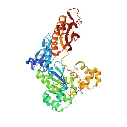
The peroxidation-derived DNA adduct, 6-oxo-M 1 dG, is a strong block to replication by human DNA polymerase eta.

Richie-Jannetta, R.Pallan, P.Kingsley, P.J.Kamdar, N.Egli, M.Marnett, L.J.

(2023) J Biological Chem 299: 105067-105067

  • DOI: https://doi.org/10.1016/j.jbc.2023.105067
  • Primary Citation Related Structures: 
    8EVE, 8EVF

  • PubMed Abstract: 

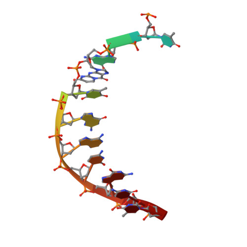
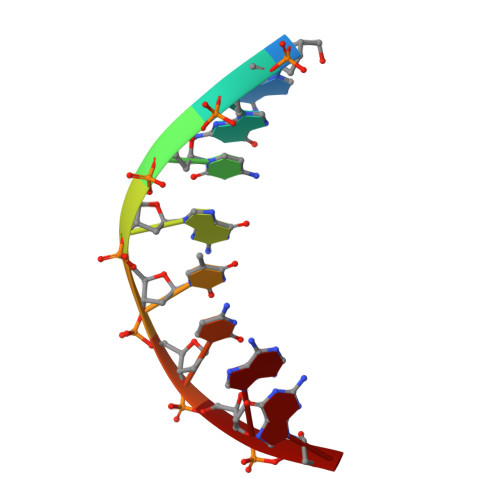
    The DNA adduct 6-oxo-M 1 dG, (3-(2'-deoxy-β-D-erythro-pentofuranosyl)-6-oxo-pyrimido(1,2alpha)purin-10(3H)-one) is formed in the genome via oxidation of the peroxidation-derived adduct M 1 dG. However, the effect of 6-oxo-M 1 dG adducts on subsequent DNA replication is unclear. Here we investigated the ability of the human Y-family polymerase hPol η to bypass 6-oxo-M 1 dG. Using steady-state kinetics and analysis of DNA extension products by liquid chromatography-tandem mass spectrometry, we found hPol η preferentially inserts a dAMP or dGMP nucleotide into primer-templates across from the 6-oxo-M 1 dG adduct, with dGMP being slightly preferred. We also show primer-templates with a 3'-terminal dGMP or dAMP across from 6-oxo-M 1 dG were extended to a greater degree than primers with a dCMP or dTMP across from the adduct. In addition, we explored the structural basis for bypass of 6-oxo-M 1 dG by hPol η using X-ray crystallography of both an insertion-stage and an extension-stage complex. In the insertion-stage complex, we observed that the incoming dCTP opposite 6-oxo-M 1 dG, although present during crystallization, was not present in the active site. We found the adduct does not interact with residues in the hPol η active site but rather forms stacking interactions with the base pair immediately 3' to the adduct. In the extension-stage complex, we observed the 3' hydroxyl group of the primer strand dGMP across from 6-oxo-M 1 dG is not positioned correctly to form a phosphodiester bond with the incoming dCTP. Taken together, these results indicate 6-oxo-M 1 dG forms a strong block to DNA replication by hPol η and provide a structural basis for its blocking ability.


  • Organizational Affiliation
    • A. B. Hancock, Jr, Memorial Laboratory for Cancer Research, Departments of Biochemistry, Chemistry and Pharmacology, Vanderbilt-Ingram Cancer Center, Vanderbilt Institute of Chemical Biology, Vanderbilt University School of Medicine, Nashville, Tennessee, USA.

Macromolecule Content 

  • Total Structure Weight: 55.35 kDa 
  • Atom Count: 3,651 
  • Modeled Residue Count: 430 
  • Deposited Residue Count: 455 
  • Unique protein chains: 1
  • Unique nucleic acid chains: 2

Macromolecules


Find similar proteins by:|  3D Structure
Entity ID: 1
MoleculeChains  Sequence LengthOrganismDetailsImage
DNA polymerase eta435Homo sapiensMutation(s): 0 
Gene Names: POLHRAD30RAD30AXPV
EC: 2.7.7.7
UniProt & NIH Common Fund Data Resources
Find proteins for Q9Y253 (Homo sapiens)
Explore Q9Y253 
Go to UniProtKB:  Q9Y253
PHAROS:  Q9Y253
GTEx:  ENSG00000170734 
Entity Groups
Sequence Clusters30% Identity50% Identity70% Identity90% Identity95% Identity100% Identity
UniProt GroupQ9Y253
Sequence Annotations
Expand
Reference Sequence
Find similar nucleic acids by:  Sequence
Entity ID: 2
MoleculeChains LengthOrganismImage
DNA 5'-D(P*TP*GP*(MO1)P*TP*GP*AP*CP*GP*CP*T-3'B [auth T]12synthetic construct
Sequence Annotations
Expand
Reference Sequence
Find similar nucleic acids by:  Sequence
Entity ID: 3
MoleculeChains LengthOrganismImage
DNA (5'-D(*AP*GP*CP*GP*TP*CP*AP*G)-3')C [auth P]8synthetic construct
Sequence Annotations
Expand
Reference Sequence

Experimental Data & Validation

Experimental Data

  • Method: X-RAY DIFFRACTION
  • Resolution: 2.87 Å
  • R-Value Free:  0.251 (Depositor), 0.251 (DCC) 
  • R-Value Work:  0.204 (Depositor), 0.204 (DCC) 
  • R-Value Observed: 0.206 (Depositor) 
Space Group: P 61
Unit Cell:
Length ( Å )Angle ( ˚ )
a = 98.671α = 90
b = 98.671β = 90
c = 81.736γ = 120
Software Package:
Software NamePurpose
HKL-2000data scaling
PHENIXrefinement
PDB_EXTRACTdata extraction
HKL-2000data reduction
MOLREPphasing

Structure Validation

View Full Validation Report


Ligand Structure Quality Assessment 


Entry History 

& Funding Information

Deposition Data


Funding OrganizationLocationGrant Number
National Institutes of Health/National Cancer Institute (NIH/NCI)United StatesR01 CA-87819
National Institutes of Health/National Cancer Institute (NIH/NCI)United StatesP01 CA-160032

Revision History  (Full details and data files)

  • Version 1.0: 2023-08-02
    Type: Initial release
  • Version 1.1: 2023-08-30
    Changes: Data collection, Database references
  • Version 1.2: 2026-03-04
    Changes: Refinement description, Structure summary