8EUN

MicroED structure of an Aeropyrum pernix protoglobin metallo-carbene complex


Experimental Data Snapshot

  • Method: ELECTRON CRYSTALLOGRAPHY
  • Resolution: 2.50 Å
  • R-Value Free: 0.284 
  • R-Value Work: 0.233 
  • R-Value Observed: 0.238 

wwPDB Validation   3D Report Full Report


This is version 1.3 of the entry. See complete history


Literature

MicroED Structure of a Protoglobin Reactive Carbene Intermediate.

Danelius, E.Porter, N.J.Unge, J.Arnold, F.H.Gonen, T.

(2023) J Am Chem Soc 145: 7159-7165

  • DOI: https://doi.org/10.1021/jacs.2c12004
  • Primary Citation of Related Structures:  
    8EUM, 8EUN

  • PubMed Abstract: 

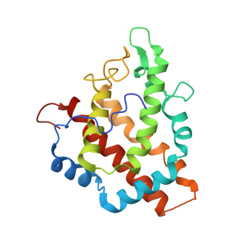
    Microcrystal electron diffraction (MicroED) is an emerging technique that has shown great potential for describing new chemical and biological molecular structures. Several important structures of small molecules, natural products, and peptides have been determined using ab initio methods. However, only a couple of novel protein structures have thus far been derived by MicroED. Taking advantage of recent technological advances, including higher acceleration voltage and using a low-noise detector in counting mode, we have determined the first structure of an Aeropyrum pernix protoglobin ( Ape Pgb) variant by MicroED using an AlphaFold2 model for phasing. The structure revealed that mutations introduced during directed evolution enhance carbene transfer activity by reorienting an α helix of Ape Pgb into a dynamic loop, making the catalytic active site more readily accessible. After exposing the tiny crystals to the substrate, we also trapped the reactive iron-carbenoid intermediate involved in this engineered Ape Pgb's new-to-nature activity, a challenging carbene transfer from a diazirine via a putative metallo-carbene. The bound structure discloses how an enlarged active site pocket stabilizes the carbene bound to the heme iron and, presumably, the transition state for the formation of this key intermediate. This work demonstrates that improved MicroED technology and the advancement in protein structure prediction now enable investigation of structures that was previously beyond reach.


  • Organizational Affiliation

    Department of Biological Chemistry, University of California, Los Angeles, 615 Charles E. Young Drive South, Los Angeles, California 90095, United States.


Macromolecules
Find similar proteins by:  (by identity cutoff)  |  3D Structure
Entity ID: 1
MoleculeChains Sequence LengthOrganismDetailsImage
Protogloblin ApPgb
A, B
195Aeropyrum pernixMutation(s): 7 
Gene Names: APE_0287
UniProt
Find proteins for Q9YFF4 (Aeropyrum pernix (strain ATCC 700893 / DSM 11879 / JCM 9820 / NBRC 100138 / K1))
Explore Q9YFF4 
Go to UniProtKB:  Q9YFF4
Entity Groups  
Sequence Clusters30% Identity50% Identity70% Identity90% Identity95% Identity100% Identity
UniProt GroupQ9YFF4
Sequence Annotations
Expand
  • Reference Sequence
Small Molecules
Ligands 1 Unique
IDChains Name / Formula / InChI Key2D Diagram3D Interactions
WUF (Subject of Investigation/LOI)
Query on WUF

Download Ideal Coordinates CCD File 
C [auth A],
D [auth B]
benzyl[3,3'-(7,12-diethenyl-3,8,13,17-tetramethylporphyrin-2,18-diyl-kappa~4~N~21~,N~22~,N~23~,N~24~)di(propanoato)(2-)]iron
C41 H39 Fe N4 O4
APVPSSILXIDIAB-HXFTUNQESA-L
Experimental Data & Validation

Experimental Data

  • Method: ELECTRON CRYSTALLOGRAPHY
  • Resolution: 2.50 Å
  • R-Value Free: 0.284 
  • R-Value Work: 0.233 
  • R-Value Observed: 0.238 
  • Space Group: P 1 21 1
Unit Cell:
Length ( Å )Angle ( ˚ )
a = 58.117α = 90
b = 45.761β = 105.72
c = 72.632γ = 90
Software Package:
Software NamePurpose
phenix.refinerefinement
PHENIXrefinement
EM Software:
TaskSoftware PackageVersion
RECONSTRUCTIONPHENIX1.20.1-4487-000

Structure Validation

View Full Validation Report


Entry History & Funding Information

Deposition Data


Funding OrganizationLocationGrant Number
National Institutes of Health/National Institute of General Medical Sciences (NIH/NIGMS)United StatesP41GM136508
Howard Hughes Medical Institute (HHMI)United States--

Revision History  (Full details and data files)

  • Version 1.0: 2023-04-05
    Type: Initial release
  • Version 1.1: 2023-04-12
    Changes: Refinement description
  • Version 1.2: 2023-04-19
    Changes: Database references
  • Version 1.3: 2024-05-22
    Changes: Data collection