Ancestral reconstruction of polyethylene terephthalate degrading cutinases reveals a rugged and unexplored sequence-fitness landscape.
Vongsouthi, V., Georgelin, R., Matthews, D.S., Saunders, J., Lee, B.M., Ton, J., Damry, A.M., Frkic, R.L., Spence, M.A., Jackson, C.J.(2025) Sci Adv 11: eads8318-eads8318
- PubMed: 40367179 Search on PubMedSearch on PubMed Central
- DOI: https://doi.org/10.1126/sciadv.ads8318
- Primary Citation Related Structures:
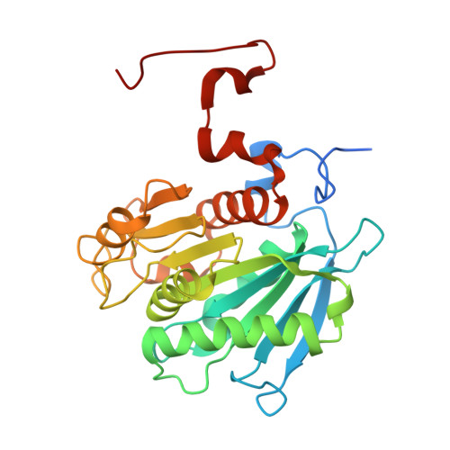
8ETX, 8ETY - PubMed Abstract:
The use of protein engineering to generate enzymes for the degradation of polyethylene terephthalate (PET) is a promising route for plastic recycling, yet traditional engineering approaches often fail to explore protein sequence space for optimal enzymes. In this work, we use multiplexed ancestral sequence reconstruction (mASR) to address this, exploring the evolutionary sequence space of PET-degrading cutinases. Using 20 statistically equivalent phylogenies of the bacterial cutinase family, we generated 48 ancestral sequences revealing a wide range of PETase activities, highlighting the value of mASR in uncovering functional variants. Our findings show PETase activity can evolve through multiple pathways involving mutations remote from the active site. Moreover, analyzing the PETase fitness landscape with local ancestral sequence embedding (LASE) revealed that LASE can capture sequence features linked to PETase activity. This work highlights mASR's potential in exploration of sequence space and underscores the use of LASE in readily mapping the protein fitness landscapes.
- Research School of Chemistry, Australian National University, Canberra, ACT 2601, Australia.
Organizational Affiliation: