8EQ5

Crystal structure of the N-terminal kinase domain of RSK2 in complex with SPRED2 (131-160)


Experimental Data Snapshot

  • Method: X-RAY DIFFRACTION
  • Resolution: 1.80 Å
  • R-Value Free: 0.225 
  • R-Value Work: 0.183 
  • R-Value Observed: 0.185 

wwPDB Validation   3D Report Full Report


Ligand Structure Quality Assessment 


This is version 1.3 of the entry. See complete history


Literature

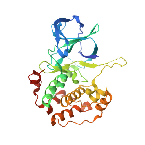
The ribosomal S6 kinase 2 (RSK2)-SPRED2 complex regulates the phosphorylation of RSK substrates and MAPK signaling.

Lopez, J.Bonsor, D.A.Sale, M.J.Urisman, A.Mehalko, J.L.Cabanski-Dunning, M.Castel, P.Simanshu, D.K.McCormick, F.

(2023) J Biol Chem 299: 104789-104789

  • DOI: https://doi.org/10.1016/j.jbc.2023.104789
  • Primary Citation of Related Structures:  
    8EQ5

  • PubMed Abstract: 

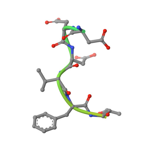
    Sprouty-related EVH-1 domain-containing (SPRED) proteins are a family of proteins that negatively regulate the RAS-Mitogen-Activated Protein Kinase (MAPK) pathway, which is involved in the regulation of the mitogenic response and cell proliferation. However, the mechanism by which these proteins affect RAS-MAPK signaling has not been elucidated. Patients with mutations in SPRED give rise to unique disease phenotypes; thus, we hypothesized that distinct interactions across SPRED proteins may account for alternative nodes of regulation. To characterize the SPRED interactome and evaluate how members of the SPRED family function through unique binding partners, we performed affinity purification mass spectrometry. We identified 90-kDa ribosomal S6 kinase 2 (RSK2) as a specific interactor of SPRED2 but not SPRED1 or SPRED3. We identified that the N-terminal kinase domain of RSK2 mediates the interaction between amino acids 123 to 201 of SPRED2. Using X-ray crystallography, we determined the structure of the SPRED2-RSK2 complex and identified the SPRED2 motif, F145A, as critical for interaction. We found that the formation of this interaction is regulated by MAPK signaling events. We also find that this interaction between SPRED2 and RSK2 has functional consequences, whereby the knockdown of SPRED2 resulted in increased phosphorylation of RSK substrates, YB1 and CREB. Furthermore, SPRED2 knockdown hindered phospho-RSK membrane and nuclear subcellular localization. We report that disruption of the SPRED2-RSK complex has effects on RAS-MAPK signaling dynamics. Our analysis reveals that members of the SPRED family have unique protein binding partners and describes the molecular and functional determinants of SPRED2-RSK2 complex dynamics.


  • Organizational Affiliation

    Helen Diller Family Comprehensive Cancer Center, University of California, San Francisco, San Francisco, California, USA.


Macromolecules
Find similar proteins by:  (by identity cutoff)  |  3D Structure
Entity ID: 1
MoleculeChains Sequence LengthOrganismDetailsImage
Ribosomal protein S6 kinase alpha-3305Homo sapiensMutation(s): 0 
Gene Names: RPS6KA3ISPK1MAPKAPK1BRSK2
EC: 2.7.11.1
UniProt & NIH Common Fund Data Resources
Find proteins for P51812 (Homo sapiens)
Explore P51812 
Go to UniProtKB:  P51812
PHAROS:  P51812
GTEx:  ENSG00000177189 
Entity Groups  
Sequence Clusters30% Identity50% Identity70% Identity90% Identity95% Identity100% Identity
UniProt GroupP51812
Sequence Annotations
Expand
  • Reference Sequence
Find similar proteins by:  (by identity cutoff)  |  3D Structure
Entity ID: 2
MoleculeChains Sequence LengthOrganismDetailsImage
Sprouty-related, EVH1 domain-containing protein 230Homo sapiensMutation(s): 0 
UniProt & NIH Common Fund Data Resources
Find proteins for Q7Z698 (Homo sapiens)
Explore Q7Z698 
Go to UniProtKB:  Q7Z698
PHAROS:  Q7Z698
GTEx:  ENSG00000198369 
Entity Groups  
Sequence Clusters30% Identity50% Identity70% Identity90% Identity95% Identity100% Identity
UniProt GroupQ7Z698
Sequence Annotations
Expand
  • Reference Sequence
Small Molecules
Ligands 1 Unique
IDChains Name / Formula / InChI Key2D Diagram3D Interactions
QCT
Query on QCT

Download Ideal Coordinates CCD File 
C [auth A]2-(3,4-dihydroxyphenyl)-5,7-dihydroxy-4-oxo-4H-chromen-3-yl 6-deoxy-alpha-L-mannopyranoside
C21 H20 O11
OXGUCUVFOIWWQJ-HQBVPOQASA-N
Experimental Data & Validation

Experimental Data

  • Method: X-RAY DIFFRACTION
  • Resolution: 1.80 Å
  • R-Value Free: 0.225 
  • R-Value Work: 0.183 
  • R-Value Observed: 0.185 
  • Space Group: I 1 2 1
Unit Cell:
Length ( Å )Angle ( ˚ )
a = 83.438α = 90
b = 41.105β = 113.87
c = 99.258γ = 90
Software Package:
Software NamePurpose
PHENIXrefinement
PDB_EXTRACTdata extraction
XDSdata reduction
Aimlessdata scaling
MOLREPphasing

Structure Validation

View Full Validation Report


Ligand Structure Quality Assessment 


Entry History & Funding Information

Deposition Data


Funding OrganizationLocationGrant Number
National Institutes of Health/National Cancer Institute (NIH/NCI)United StatesHHSN261200800001E

Revision History  (Full details and data files)

  • Version 1.0: 2023-04-26
    Type: Initial release
  • Version 1.1: 2023-05-24
    Changes: Database references
  • Version 1.2: 2023-06-14
    Changes: Database references
  • Version 1.3: 2023-10-25
    Changes: Data collection, Refinement description