


Crystal Structure of the Beta-lactamase Class D from Chitinophaga pinensis in the complex with Avibactam.
Maltseva, N., Kim, Y., Endres, M., Joachimiak, A.To be published.
Experimental Data Snapshot
Entity ID: 1 | |||||
|---|---|---|---|---|---|
| Molecule | Chains | Sequence Length | Organism | Details | Image |
| Beta-lactamase Class D Cpin_0907 | 247 | Chitinophaga pinensis DSM 2588 | Mutation(s): 0 Gene Names: blaOXA, FEF09_01595 | 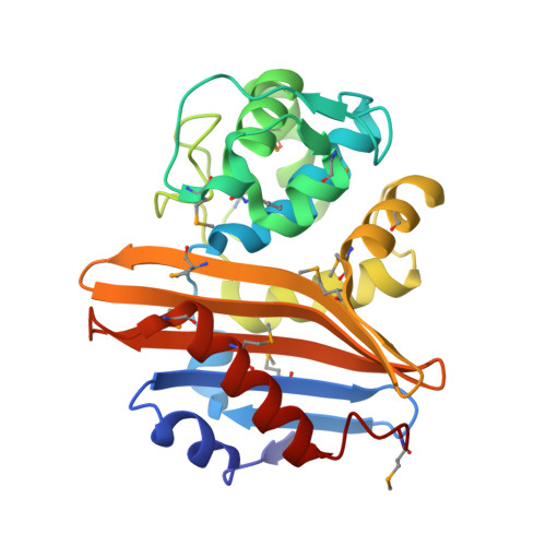 | |
UniProt | |||||
Entity Groups | |||||
| Sequence Clusters | 30% Identity50% Identity70% Identity90% Identity95% Identity100% Identity | ||||
| UniProt Group | A0A5C6M0R4 | ||||
Sequence AnnotationsExpand | |||||
Reference Sequence | |||||
| Ligands 2 Unique | |||||
|---|---|---|---|---|---|
| ID | Chains | Name / Formula / InChI Key | 2D Diagram | 3D Interactions | |
| NXL (Subject of Investigation/LOI) Download:Ideal Coordinates CCD File | B [auth A] | (2S,5R)-1-formyl-5-[(sulfooxy)amino]piperidine-2-carboxamide C7 H13 N3 O6 S WJDGWXPPFHLLNL-RITPCOANSA-N |  | ||
| ACY Download:Ideal Coordinates CCD File | C [auth A] | ACETIC ACID C2 H4 O2 QTBSBXVTEAMEQO-UHFFFAOYSA-N |  | ||
| Modified Residues 2 Unique | |||||
|---|---|---|---|---|---|
| ID | Chains | Type | Formula | 2D Diagram | Parent |
| KCX Query on KCX | A | L-PEPTIDE LINKING | C7 H14 N2 O4 |  | LYS |
| MSE Query on MSE | A | L-PEPTIDE LINKING | C5 H11 N O2 Se |  | MET |
| Length ( Å ) | Angle ( ˚ ) |
|---|---|
| a = 48.816 | α = 90 |
| b = 67.864 | β = 90 |
| c = 68.698 | γ = 90 |
| Software Name | Purpose |
|---|---|
| PHENIX | refinement |
| HKL-3000 | data reduction |
| HKL-3000 | data scaling |
| HKL-3000 | phasing |
| MLPHARE | phasing |
| Funding Organization | Location | Grant Number |
|---|---|---|
| National Institutes of Health/National Institute Of Allergy and Infectious Diseases (NIH/NIAID) | United States | -- |


