Precisely patterned nanofibres made from extendable protein multiplexes.
Bethel, N.P., Borst, A.J., Parmeggiani, F., Bick, M.J., Brunette, T.J., Nguyen, H., Kang, A., Bera, A.K., Carter, L., Miranda, M.C., Kibler, R.D., Lamb, M., Li, X., Sankaran, B., Baker, D.(2023) Nat Chem 15: 1664-1671
- PubMed: 37667012 Search on PubMedSearch on PubMed Central
- DOI: https://doi.org/10.1038/s41557-023-01314-x
- Primary Citation Related Structures:
8EOV, 8EOX, 8EOZ, 8ERW, 8G8I, 8GA9, 8GAA, 8GAQ - PubMed Abstract:
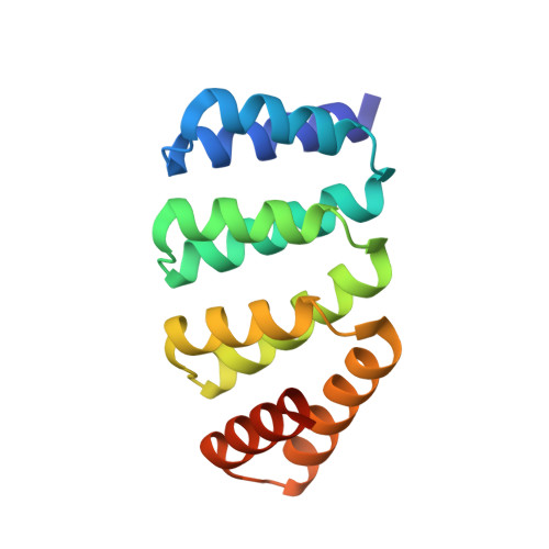
Molecular systems with coincident cyclic and superhelical symmetry axes have considerable advantages for materials design as they can be readily lengthened or shortened by changing the length of the constituent monomers. Among proteins, alpha-helical coiled coils have such symmetric, extendable architectures, but are limited by the relatively fixed geometry and flexibility of the helical protomers. Here we describe a systematic approach to generating modular and rigid repeat protein oligomers with coincident C 2 to C 8 and superhelical symmetry axes that can be readily extended by repeat propagation. From these building blocks, we demonstrate that a wide range of unbounded fibres can be systematically designed by introducing hydrophilic surface patches that force staggering of the monomers; the geometry of such fibres can be precisely tuned by varying the number of repeat units in the monomer and the placement of the hydrophilic patches.
- Department of Biochemistry, University of Washington, Seattle, WA, USA.
Organizational Affiliation: