8END

Crystal structure of LGR ligand alpha2/beta5 from C. elegans in crystal form 1 (SeMet)


Experimental Data Snapshot

  • Method: X-RAY DIFFRACTION
  • Resolution: 3.54 Å
  • R-Value Free: 0.330 
  • R-Value Work: 0.282 
  • R-Value Observed: 0.286 

wwPDB Validation   3D Report Full Report


This is version 1.3 of the entry. See complete history


Literature

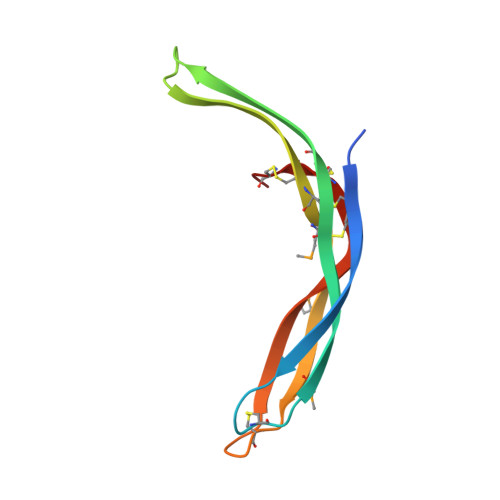
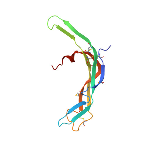
Crystal structure of LGR ligand alpha2/beta5 from Caenorhabditis elegans with implications for the evolution of glycoprotein hormones

Gong, Z.Wang, W.El Omari, K.Lebedev, A.A.Clarke, O.B.Hendrickson, W.A.

(2023) Proc Natl Acad Sci U S A 120: e2218630120

  • DOI: https://doi.org/10.1073/pnas.2218630120
  • Primary Citation of Related Structures:  
    8ENB, 8END, 8ENF

  • PubMed Abstract: 

    A family of leucine-rich-repeat-containing G-protein-coupled receptors (LGRs) mediate diverse physiological responses when complexed with their cognate ligands. LGRs are present in all metazoan animals. In humans, the LGR ligands include glycoprotein hormones (GPHs) chorionic gonadotropin (hCG), luteinizing hormone, follicle-stimulating hormone (hFSH), and thyroid-stimulating hormone (hTSH). These hormones are αβ heterodimers of cystine-knot protein chains. LGRs and their ligand chains have coevolved. Ancestral hormone homologs, present in both bilaterian animals and chordates, are identified as α2β5. We have used single-wavelength anomalous diffraction and molecular replacement to determine structures of the α2β5 hormone from Caenorhabditis elegans ( Ce α2β5). Ce α2β5 is unglycosylated, as are many other α2β5 hormones. Both Hs α2β5, the human homolog of Ce α2β5, and hTSH activate the same receptor (hTSHR). Despite having little sequence similarity to vertebrate GPHs, apart from the cysteine patterns from core disulfide bridges, Ce α2β5 is generally similar in structure to these counterparts; however, its α2 and β5 subunits are more symmetric as compared with α and β of hCG and hFSH. This quasisymmetry suggests a hypothetical homodimeric antecedent of the α2β5 and αβ heterodimers. Known structures together with AlphaFold models from the sequences for other LGR ligands provide representatives for the molecular evolution of LGR ligands from early metazoans through the present-day GPHs. The experimental Ce α2β5 structure validates its AlphaFold model, and thus also that for Hs α2β5; and interfacial characteristics in a model for the Hs α2β5:hTSHR complex are similar to those found in an experimental hTSH:hTSHR structure.


  • Organizational Affiliation

    Department of Biochemistry and Molecular Biophysics, Columbia University, New York, NY 10032.


Macromolecules
Find similar proteins by:  (by identity cutoff)  |  3D Structure
Entity ID: 1
MoleculeChains Sequence LengthOrganismDetailsImage
Bursicon92Caenorhabditis elegansMutation(s): 0 
UniProt
Find proteins for A0T3A2 (Caenorhabditis elegans)
Explore A0T3A2 
Go to UniProtKB:  A0T3A2
Entity Groups  
Sequence Clusters30% Identity50% Identity70% Identity90% Identity95% Identity100% Identity
UniProt GroupA0T3A2
Sequence Annotations
Expand
  • Reference Sequence
Find similar proteins by:  (by identity cutoff)  |  3D Structure
Entity ID: 2
MoleculeChains Sequence LengthOrganismDetailsImage
Cys_knot domain-containing protein106Caenorhabditis elegansMutation(s): 0 
Gene Names: gpb5CELE_T23B12.8T23B12.8
UniProt
Find proteins for A7DT38 (Caenorhabditis elegans)
Explore A7DT38 
Go to UniProtKB:  A7DT38
Entity Groups  
Sequence Clusters30% Identity50% Identity70% Identity90% Identity95% Identity100% Identity
UniProt GroupA7DT38
Sequence Annotations
Expand
  • Reference Sequence
Small Molecules
Modified Residues  1 Unique
IDChains TypeFormula2D DiagramParent
MSE
Query on MSE
A
L-PEPTIDE LINKINGC5 H11 N O2 SeMET
Experimental Data & Validation

Experimental Data

  • Method: X-RAY DIFFRACTION
  • Resolution: 3.54 Å
  • R-Value Free: 0.330 
  • R-Value Work: 0.282 
  • R-Value Observed: 0.286 
  • Space Group: P 32 2 1
Unit Cell:
Length ( Å )Angle ( ˚ )
a = 92.53α = 90
b = 92.53β = 90
c = 95.6γ = 120
Software Package:
Software NamePurpose
PHENIXrefinement
XDSdata reduction
STARANISOdata scaling
Cootmodel building

Structure Validation

View Full Validation Report


Entry History & Funding Information

Deposition Data


Funding OrganizationLocationGrant Number
National Institutes of Health/National Institute of General Medical Sciences (NIH/NIGMS)United StatesGM107462

Revision History  (Full details and data files)

  • Version 1.0: 2023-01-11
    Type: Initial release
  • Version 1.1: 2023-04-05
    Changes: Refinement description, Structure summary
  • Version 1.2: 2023-10-25
    Changes: Data collection, Refinement description
  • Version 1.3: 2023-11-15
    Changes: Data collection