De novo designed ice-binding proteins from twist-constrained helices.
de Haas, R.J., Tas, R.P., van den Broek, D., Zheng, C., Nguyen, H., Kang, A., Bera, A.K., King, N.P., Voets, I.K., de Vries, R.(2023) Proc Natl Acad Sci U S A 120: e2220380120-e2220380120
- PubMed: 37364125 Search on PubMedSearch on PubMed Central
- DOI: https://doi.org/10.1073/pnas.2220380120
- Primary Citation Related Structures:
8EK4 - PubMed Abstract:
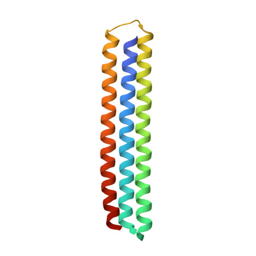
Attaining molecular-level control over solidification processes is a crucial aspect of materials science. To control ice formation, organisms have evolved bewildering arrays of ice-binding proteins (IBPs), but these have poorly understood structure-activity relationships. We propose that reverse engineering using de novo computational protein design can shed light on structure-activity relationships of IBPs. We hypothesized that the model alpha-helical winter flounder antifreeze protein uses an unusual undertwisting of its alpha-helix to align its putative ice-binding threonine residues in exactly the same direction. We test this hypothesis by designing a series of straight three-helix bundles with an ice-binding helix projecting threonines and two supporting helices constraining the twist of the ice-binding helix. Our findings show that ice-recrystallization inhibition by the designed proteins increases with the degree of designed undertwisting, thus validating our hypothesis, and opening up avenues for the computational design of IBPs.
- Department of Physical Chemistry and Soft Matter, Wageningen University and Research, Wageningen, WE 6708, The Netherlands.
Organizational Affiliation: