Mitoguardin-2-mediated lipid transfer preserves mitochondrial morphology and lipid droplet formation.
Hong, Z., Adlakha, J., Wan, N., Guinn, E., Giska, F., Gupta, K., Melia, T.J., Reinisch, K.M.(2022) J Cell Biol 221
- PubMed: 36282247 Search on PubMedSearch on PubMed Central
- DOI: https://doi.org/10.1083/jcb.202207022
- Primary Citation Related Structures:
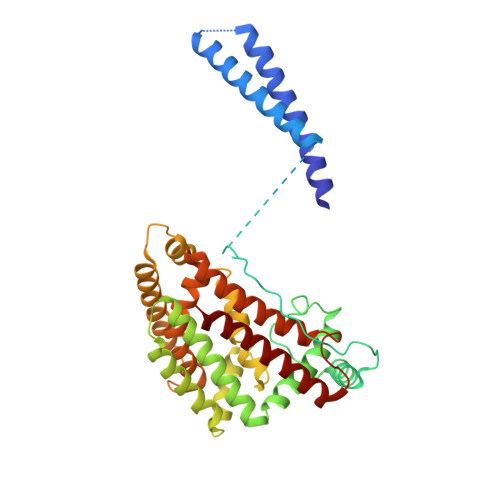
8EDV - PubMed Abstract:
Lipid transport proteins at membrane contacts, where organelles are closely apposed, are critical in redistributing lipids from the endoplasmic reticulum (ER), where they are made, to other cellular membranes. Such protein-mediated transfer is especially important for maintaining organelles disconnected from secretory pathways, like mitochondria. We identify mitoguardin-2, a mitochondrial protein at contacts with the ER and/or lipid droplets (LDs), as a lipid transporter. An x-ray structure shows that the C-terminal domain of mitoguardin-2 has a hydrophobic cavity that binds lipids. Mass spectrometry analysis reveals that both glycerophospholipids and free-fatty acids co-purify with mitoguardin-2 from cells, and that each mitoguardin-2 can accommodate up to two lipids. Mitoguardin-2 transfers glycerophospholipids between membranes in vitro, and this transport ability is required for roles both in mitochondrial and LD biology. While it is not established that protein-mediated transfer at contacts plays a role in LD metabolism, our findings raise the possibility that mitoguardin-2 functions in transporting fatty acids and glycerophospholipids at mitochondria-LD contacts.
- Department of Cell Biology, Yale University School of Medicine, New Haven, CT.
Organizational Affiliation: