8ED4

Structure of the complex between the arsenite oxidase and its native electron acceptor cytochrome c552 from Pseudorhizobium sp. str. NT-26


Experimental Data Snapshot

  • Method: X-RAY DIFFRACTION
  • Resolution: 2.25 Å
  • R-Value Free: 0.230 
  • R-Value Work: 0.181 
  • R-Value Observed: 0.183 

wwPDB Validation   3D Report Full Report


Ligand Structure Quality Assessment 


This is version 1.1 of the entry. See complete history


Literature

The structure of the complex between the arsenite oxidase from Pseudorhizobium banfieldiae sp. strain NT-26 and its native electron acceptor cytochrome c 552.

Poddar, N.Santini, J.M.Maher, M.J.

(2023) Acta Crystallogr D Struct Biol 79: 345-352

  • DOI: https://doi.org/10.1107/S2059798323002103
  • Primary Citation of Related Structures:  
    8ED4

  • PubMed Abstract: 

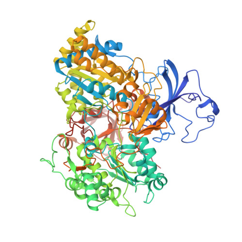
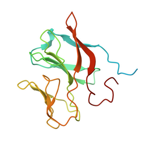
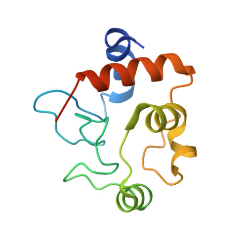
    The arsenite oxidase (AioAB) from Pseudorhizobium banfieldiae sp. strain NT-26 catalyzes the oxidation of arsenite to arsenate and transfers electrons to its cognate electron acceptor cytochrome c 552 (cytc 552 ). This activity underpins the ability of this organism to respire using arsenite present in contaminated environments. The crystal structure of the AioAB/cytc 552 electron transfer complex reveals two A 2 B 2 /(cytc 552 ) 2 assemblies per asymmetric unit. Three of the four cytc 552 molecules in the asymmetric unit dock to AioAB in a cleft at the interface between the AioA and AioB subunits, with an edge-to-edge distance of 7.5 Å between the heme of cytc 552 and the [2Fe-2S] Rieske cluster in the AioB subunit. The interface between the AioAB and cytc 552 proteins features electrostatic and nonpolar interactions and is stabilized by two salt bridges. A modest number of hydrogen bonds, salt bridges and relatively small, buried surface areas between protein partners are typical features of transient electron transfer complexes. Interestingly, the fourth cytc 552 molecule is positioned differently between two AioAB heterodimers, with distances between its heme and the AioAB redox active cofactors that are outside the acceptable range for fast electron transfer. This unique cytc 552 molecule appears to be positioned to facilitate crystal packing rather than reflecting a functional complex.


  • Organizational Affiliation

    School of Chemistry and The Bio21 Molecular Science and Biotechnology Institute, The University of Melbourne, Parkville, Australia.


Macromolecules
Find similar proteins by:  (by identity cutoff)  |  3D Structure
Entity ID: 1
MoleculeChains Sequence LengthOrganismDetailsImage
AroAA,
D [auth C],
G [auth E],
J [auth G]
844Pseudorhizobium banfieldiaeMutation(s): 0 
Gene Names: aroAaioANT26_p10030
EC: 1.20.98.1
UniProt
Find proteins for Q6VAL8 (Pseudorhizobium banfieldiae)
Explore Q6VAL8 
Go to UniProtKB:  Q6VAL8
Entity Groups  
Sequence Clusters30% Identity50% Identity70% Identity90% Identity95% Identity100% Identity
UniProt GroupQ6VAL8
Sequence Annotations
Expand
  • Reference Sequence
Find similar proteins by:  (by identity cutoff)  |  3D Structure
Entity ID: 2
MoleculeChains Sequence LengthOrganismDetailsImage
AroBB,
E [auth D],
H [auth F],
K [auth H]
162Pseudorhizobium banfieldiaeMutation(s): 0 
Gene Names: aroB
UniProt
Find proteins for Q6VAL9 (Pseudorhizobium banfieldiae)
Explore Q6VAL9 
Go to UniProtKB:  Q6VAL9
Entity Groups  
Sequence Clusters30% Identity50% Identity70% Identity90% Identity95% Identity100% Identity
UniProt GroupQ6VAL9
Sequence Annotations
Expand
  • Reference Sequence
Find similar proteins by:  (by identity cutoff)  |  3D Structure
Entity ID: 3
MoleculeChains Sequence LengthOrganismDetailsImage
C-type cytochrome c552C [auth I],
F [auth J],
I [auth K],
L
117Pseudorhizobium banfieldiaeMutation(s): 0 
Gene Names: cytCNT26_p10031
UniProt
Find proteins for Q2TV05 (Pseudorhizobium banfieldiae)
Explore Q2TV05 
Go to UniProtKB:  Q2TV05
Entity Groups  
Sequence Clusters30% Identity50% Identity70% Identity90% Identity95% Identity100% Identity
UniProt GroupQ2TV05
Sequence Annotations
Expand
  • Reference Sequence
Small Molecules
Ligands 9 Unique
IDChains Name / Formula / InChI Key2D Diagram3D Interactions
MGD
Query on MGD

Download Ideal Coordinates CCD File 
AA [auth E]
BA [auth E]
JA [auth G]
KA [auth G]
M [auth A]
AA [auth E],
BA [auth E],
JA [auth G],
KA [auth G],
M [auth A],
N [auth A],
T [auth C],
U [auth C]
2-AMINO-5,6-DIMERCAPTO-7-METHYL-3,7,8A,9-TETRAHYDRO-8-OXA-1,3,9,10-TETRAAZA-ANTHRACEN-4-ONE GUANOSINE DINUCLEOTIDE
C20 H26 N10 O13 P2 S2
VQAGYJCYOLHZDH-ILXWUORBSA-N
HEC
Query on HEC

Download Ideal Coordinates CCD File 
IA [auth K]HEME C
C34 H34 Fe N4 O4
HXQIYSLZKNYNMH-LJNAALQVSA-N
HEM
Query on HEM

Download Ideal Coordinates CCD File 
QA [auth L],
S [auth I],
Z [auth J]
PROTOPORPHYRIN IX CONTAINING FE
C34 H32 Fe N4 O4
KABFMIBPWCXCRK-RGGAHWMASA-L
F3S
Query on F3S

Download Ideal Coordinates CCD File 
DA [auth E],
MA [auth G],
P [auth A],
W [auth C]
FE3-S4 CLUSTER
Fe3 S4
FCXHZBQOKRZXKS-UHFFFAOYSA-N
PG4
Query on PG4

Download Ideal Coordinates CCD File 
GA [auth E],
OA [auth G]
TETRAETHYLENE GLYCOL
C8 H18 O5
UWHCKJMYHZGTIT-UHFFFAOYSA-N
FES
Query on FES

Download Ideal Coordinates CCD File 
HA [auth F],
PA [auth H],
R [auth B],
Y [auth D]
FE2/S2 (INORGANIC) CLUSTER
Fe2 S2
NIXDOXVAJZFRNF-UHFFFAOYSA-N
4MO
Query on 4MO

Download Ideal Coordinates CCD File 
CA [auth E],
LA [auth G],
O [auth A],
V [auth C]
MOLYBDENUM(IV) ION
Mo
ZIKKVZAYJJZBGE-UHFFFAOYSA-N
GOL
Query on GOL

Download Ideal Coordinates CCD File 
FA [auth E]GLYCEROL
C3 H8 O3
PEDCQBHIVMGVHV-UHFFFAOYSA-N
O
Query on O

Download Ideal Coordinates CCD File 
EA [auth E],
NA [auth G],
Q [auth A],
X [auth C]
OXYGEN ATOM
O
XLYOFNOQVPJJNP-UHFFFAOYSA-N
Experimental Data & Validation

Experimental Data

  • Method: X-RAY DIFFRACTION
  • Resolution: 2.25 Å
  • R-Value Free: 0.230 
  • R-Value Work: 0.181 
  • R-Value Observed: 0.183 
  • Space Group: P 1 21 1
Unit Cell:
Length ( Å )Angle ( ˚ )
a = 129.4α = 90
b = 126.56β = 107.81
c = 148.02γ = 90
Software Package:
Software NamePurpose
REFMACrefinement
PDB_EXTRACTdata extraction
XDSdata reduction
Aimlessdata scaling
PHASERphasing

Structure Validation

View Full Validation Report


Ligand Structure Quality Assessment 


Entry History & Funding Information

Deposition Data


Funding OrganizationLocationGrant Number
Not funded--

Revision History  (Full details and data files)

  • Version 1.0: 2023-04-12
    Type: Initial release
  • Version 1.1: 2023-10-25
    Changes: Data collection, Refinement description