8E50

Cryo-EM structure of human glycerol-3-phosphate acyltransferase 1 (GPAT1) in complex with CoA and palmitoyl-LPA


Experimental Data Snapshot

  • Method: ELECTRON MICROSCOPY
  • Resolution: 3.67 Å
  • Aggregation State: PARTICLE 
  • Reconstruction Method: SINGLE PARTICLE 

wwPDB Validation   3D Report Full Report


This is version 1.2 of the entry. See complete history


Literature

Structural basis of the acyl-transfer mechanism of human GPAT1.

Johnson, Z.L.Ammirati, M.Wasilko, D.J.Chang, J.S.Noell, S.Foley, T.L.Yoon, H.Smith, K.Asano, S.Hales, K.Wan, M.Yang, Q.Piotrowski, M.A.Farley, K.A.Gilbert, T.Aschenbrenner, L.M.Fennell, K.F.Dutra, J.K.Xu, M.Guo, C.Varghese, A.E.Bellenger, J.Quinn, A.Am Ende, C.W.West, G.M.Griffor, M.C.Bennett, D.Calabrese, M.Steppan, C.M.Han, S.Wu, H.

(2023) Nat Struct Mol Biol 30: 22-30

  • DOI: https://doi.org/10.1038/s41594-022-00884-7
  • Primary Citation of Related Structures:  
    8E4Y, 8E50

  • PubMed Abstract: 

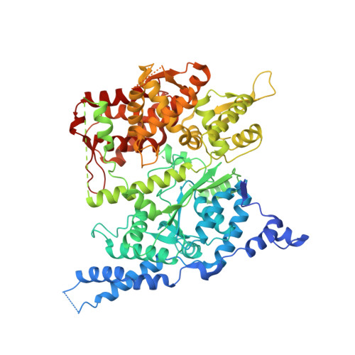
    Glycerol-3-phosphate acyltransferase (GPAT)1 is a mitochondrial outer membrane protein that catalyzes the first step of de novo glycerolipid biosynthesis. Hepatic expression of GPAT1 is linked to liver fat accumulation and the severity of nonalcoholic fatty liver diseases. Here we present the cryo-EM structures of human GPAT1 in substrate analog-bound and product-bound states. The structures reveal an N-terminal acyltransferase domain that harbors important catalytic motifs and a tightly associated C-terminal domain that is critical for proper protein folding. Unexpectedly, GPAT1 has no transmembrane regions as previously proposed but instead associates with the membrane via an amphipathic surface patch and an N-terminal loop-helix region that contains a mitochondrial-targeting signal. Combined structural, computational and functional studies uncover a hydrophobic pathway within GPAT1 for lipid trafficking. The results presented herein lay a framework for rational inhibitor development for GPAT1.


  • Organizational Affiliation

    Discovery Sciences, Medicine Design, Pfizer Inc., Groton, CT, USA.


Macromolecules
Find similar proteins by:  (by identity cutoff)  |  3D Structure
Entity ID: 1
MoleculeChains Sequence LengthOrganismDetailsImage
Glycerol-3-phosphate acyltransferase 1, mitochondrial767Homo sapiensMutation(s): 0 
Gene Names: GPAMGPAT1KIAA1560
EC: 2.3.1.15
UniProt & NIH Common Fund Data Resources
Find proteins for Q9HCL2 (Homo sapiens)
Explore Q9HCL2 
Go to UniProtKB:  Q9HCL2
PHAROS:  Q9HCL2
GTEx:  ENSG00000119927 
Entity Groups  
Sequence Clusters30% Identity50% Identity70% Identity90% Identity95% Identity100% Identity
UniProt GroupQ9HCL2
Sequence Annotations
Expand
  • Reference Sequence
Small Molecules
Ligands 2 Unique
IDChains Name / Formula / InChI Key2D Diagram3D Interactions
COA (Subject of Investigation/LOI)
Query on COA

Download Ideal Coordinates CCD File 
B [auth A]COENZYME A
C21 H36 N7 O16 P3 S
RGJOEKWQDUBAIZ-IBOSZNHHSA-N
NKO (Subject of Investigation/LOI)
Query on NKO

Download Ideal Coordinates CCD File 
C [auth A](2R)-2-hydroxy-3-(phosphonooxy)propyl hexadecanoate
C19 H39 O7 P
YNDYKPRNFWPPFU-GOSISDBHSA-N
Experimental Data & Validation

Experimental Data

  • Method: ELECTRON MICROSCOPY
  • Resolution: 3.67 Å
  • Aggregation State: PARTICLE 
  • Reconstruction Method: SINGLE PARTICLE 
EM Software:
TaskSoftware PackageVersion
RECONSTRUCTIONRELION3.1.2
MODEL REFINEMENTPHENIX
MODEL REFINEMENTREFMAC5

Structure Validation

View Full Validation Report


Entry History & Funding Information

Deposition Data


Funding OrganizationLocationGrant Number
Not funded--

Revision History  (Full details and data files)

  • Version 1.0: 2022-12-21
    Type: Initial release
  • Version 1.1: 2022-12-28
    Changes: Database references
  • Version 1.2: 2023-02-01
    Changes: Database references