Crystal Structure of CYP3A4 Complexed with Fluorol Identifies the Substrate Access Channel as a High-Affinity Ligand Binding Site.
Sevrioukova, I.F.(2022) Int J Mol Sci 23
- PubMed: 36293445 Search on PubMedSearch on PubMed Central
- DOI: https://doi.org/10.3390/ijms232012591
- Primary Citation Related Structures:
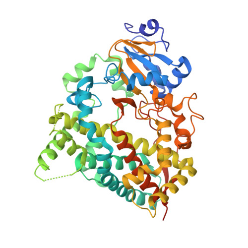
8DYC - PubMed Abstract:
Cytochrome P450 3A4 (CYP3A4) is a major human drug-metabolizing enzyme, notoriously known for its extreme substrate promiscuity, allosteric behavior, and implications in drug-drug interactions. Despite extensive investigations, the mechanism of ligand binding to CYP3A4 is not fully understood. We determined the crystal structure of CYP3A4 complexed with fluorol, a small fluorescent dye that can undergo hydroxylation. In the structure, fluorol associates to the substrate channel, well suited for the binding of planar polyaromatic molecules bearing polar groups, through which stabilizing H-bonds with the polar channel residues, such as Thr224 and Arg372, can be established. Mutagenesis, spectral, kinetic, and functional data confirmed the involvement but not strict requirement of Thr224 for the association of fluorol. Collectively, our data identify the substrate channel as a high-affinity ligand binding site and support the notion that hydrophobic ligands first dock to the nearby peripheral surface, before migrating to the channel and, subsequently, into the active site.
- Department of Molecular Biology and Biochemistry, University of California, Irvine, CA 92697-3900, USA.
Organizational Affiliation: