


Crystal Structure of a Putative D-beta-hydroxybutyrate dehydrogenase from Burkholderia cenocepacia J2315 in complex with NAD
Abendroth, J., Mayclin, S.J., Phan, J.N., Lorimer, D.D., Horanyi, P.S., Edwards, T.E.To be published.
Experimental Data Snapshot
Starting Model: experimental
View more details
Entity ID: 1 | |||||
|---|---|---|---|---|---|
| Molecule | Chains | Sequence Length | Organism | Details | Image |
| 3-hydroxybutyrate dehydrogenase | 269 | Burkholderia cenocepacia | Mutation(s): 0 Gene Names: A8E72_04365, A8F33_24625 | 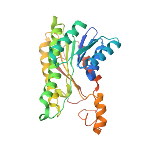 | |
UniProt | |||||
Find proteins for A0A1V2Y0M0 (Burkholderia cenocepacia) Explore A0A1V2Y0M0 Go to UniProtKB: A0A1V2Y0M0 | |||||
Entity Groups | |||||
| Sequence Clusters | 30% Identity50% Identity70% Identity90% Identity95% Identity100% Identity | ||||
| UniProt Group | A0A1V2Y0M0 | ||||
Sequence AnnotationsExpand | |||||
Reference Sequence | |||||
| Ligands 4 Unique | |||||
|---|---|---|---|---|---|
| ID | Chains | Name / Formula / InChI Key | 2D Diagram | 3D Interactions | |
| NAD (Subject of Investigation/LOI) Download:Ideal Coordinates CCD File | E [auth A], J [auth B], N [auth C], R [auth D] | NICOTINAMIDE-ADENINE-DINUCLEOTIDE C21 H27 N7 O14 P2 BAWFJGJZGIEFAR-NNYOXOHSSA-N |  | ||
| PG4 Download:Ideal Coordinates CCD File | H [auth A] I [auth A] L [auth B] M [auth B] P [auth C] | TETRAETHYLENE GLYCOL C8 H18 O5 UWHCKJMYHZGTIT-UHFFFAOYSA-N |  | ||
| CIT Download:Ideal Coordinates CCD File | F [auth A], K [auth B], O [auth C], S [auth D] | CITRIC ACID C6 H8 O7 KRKNYBCHXYNGOX-UHFFFAOYSA-N |  | ||
| PO4 Download:Ideal Coordinates CCD File | G [auth A] | PHOSPHATE ION O4 P NBIIXXVUZAFLBC-UHFFFAOYSA-K |  | ||
| Length ( Å ) | Angle ( ˚ ) |
|---|---|
| a = 59.91 | α = 97.149 |
| b = 65.6 | β = 98.382 |
| c = 68.32 | γ = 107.47 |
| Software Name | Purpose |
|---|---|
| XDS | data reduction |
| XSCALE | data scaling |
| PHENIX | refinement |
| PDB_EXTRACT | data extraction |
| PHASER | phasing |
| Funding Organization | Location | Grant Number |
|---|---|---|
| National Institutes of Health/National Institute Of Allergy and Infectious Diseases (NIH/NIAID) | United States | HHSN272201700059C |


