Scaffolding protein functional sites using deep learning.
Wang, J., Lisanza, S., Juergens, D., Tischer, D., Watson, J.L., Castro, K.M., Ragotte, R., Saragovi, A., Milles, L.F., Baek, M., Anishchenko, I., Yang, W., Hicks, D.R., Exposit, M., Schlichthaerle, T., Chun, J.H., Dauparas, J., Bennett, N., Wicky, B.I.M., Muenks, A., DiMaio, F., Correia, B., Ovchinnikov, S., Baker, D.(2022) Science 377: 387-394
- PubMed: 35862514 Search on PubMedSearch on PubMed Central
- DOI: https://doi.org/10.1126/science.abn2100
- Primary Citation Related Structures:
8DT0 - PubMed Abstract:
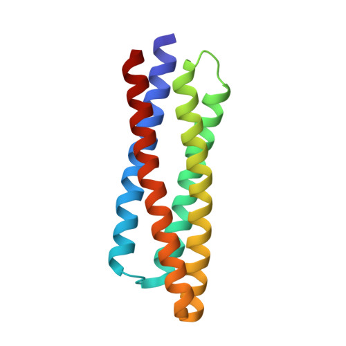
The binding and catalytic functions of proteins are generally mediated by a small number of functional residues held in place by the overall protein structure. Here, we describe deep learning approaches for scaffolding such functional sites without needing to prespecify the fold or secondary structure of the scaffold. The first approach, "constrained hallucination," optimizes sequences such that their predicted structures contain the desired functional site. The second approach, "inpainting," starts from the functional site and fills in additional sequence and structure to create a viable protein scaffold in a single forward pass through a specifically trained RoseTTAFold network. We use these two methods to design candidate immunogens, receptor traps, metalloproteins, enzymes, and protein-binding proteins and validate the designs using a combination of in silico and experimental tests.
- Department of Biochemistry, University of Washington, Seattle, WA 98105, USA.
Organizational Affiliation: