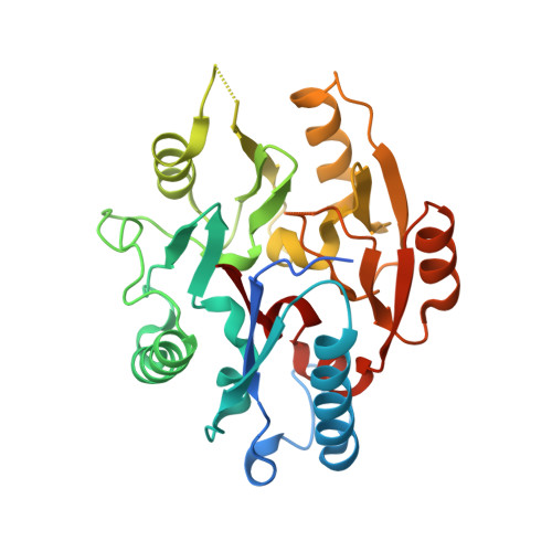
Structure of the T. brucei kinetoplastid RNA editing substrate-binding complex core component, RESC5.
Salinas, R., Cannistraci, E., Schumacher, M.A.(2023) PLoS One 18: e0282155-e0282155
- PubMed: 36862634 Search on PubMedSearch on PubMed Central
- DOI: https://doi.org/10.1371/journal.pone.0282155
- Primary Citation Related Structures:
8DPK - PubMed Abstract:
Kinetoplastid protists such as Trypanosoma brucei undergo an unusual process of mitochondrial uridine (U) insertion and deletion editing termed kinetoplastid RNA editing (kRNA editing). This extensive form of editing, which is mediated by guide RNAs (gRNAs), can involve the insertion of hundreds of Us and deletion of tens of Us to form a functional mitochondrial mRNA transcript. kRNA editing is catalyzed by the 20 S editosome/RECC. However, gRNA directed, processive editing requires the RNA editing substrate binding complex (RESC), which is comprised of 6 core proteins, RESC1-RESC6. To date there are no structures of RESC proteins or complexes and because RESC proteins show no homology to proteins of known structure, their molecular architecture remains unknown. RESC5 is a key core component in forming the foundation of the RESC complex. To gain insight into the RESC5 protein we performed biochemical and structural studies. We show that RESC5 is monomeric and we report the T. brucei RESC5 crystal structure to 1.95 Å. RESC5 harbors a dimethylarginine dimethylaminohydrolase-like (DDAH) fold. DDAH enzymes hydrolyze methylated arginine residues produced during protein degradation. However, RESC5 is missing two key catalytic DDAH residues and does bind DDAH substrate or product. Implications of the fold for RESC5 function are discussed. This structure provides the first structural view of an RESC protein.
- Department of Biochemistry, Duke University School of Medicine, DUMC, Durham, NC, United States of America.
Organizational Affiliation: