Ibuprofen: a weak inhibitor of carbonic anhydrase II.
Combs, J., Andring, J., McKenna, R.(2022) Acta Crystallogr F Struct Biol Commun 78: 395-402
- PubMed: 36322425 Search on PubMedSearch on PubMed Central
- DOI: https://doi.org/10.1107/S2053230X22009761
- Primary Citation Related Structures:
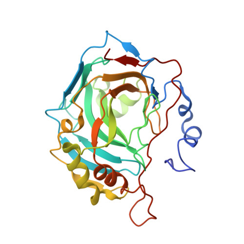
8DJ9 - PubMed Abstract:
Carbonic anhydrases (CAs) are drug targets for a variety of diseases. While many clinically relevant CA inhibitors are sulfonamide-based, novel CA inhibitors are being developed that incorporate alternative zinc-binding groups, such as carboxylic acid moieties, to develop CA isoform-specific inhibitors. Here, the X-ray crystal structure of human CA II (hCA II) in complex with the carboxylic acid ibuprofen [2-(4-isobutylphenyl)propanoic acid, a common over-the-counter nonsteroidal anti-inflammatory drug] is reported to 1.54 Å resolution. The binding of ibuprofen is overlaid with the structures of other carboxylic acids in complex with hCA II to compare their inhibition mechanisms by direct or indirect (via a water) binding to the active-site zinc. Additionally, enzyme-inhibition assays using ibuprofen, nicotinic acid and ferulic acid were performed with hCA II to determine their IC 50 values and were compared with those of other carboxylic acid binders. This study discusses the potential development of CA inhibitors utilizing the carboxylic acid moiety.
- Department of Biochemistry and Molecular Biology, College of Medicine, University of Florida, Gainesville, FL 32610, USA.
Organizational Affiliation: