


Structure and Function of Engineered Variants of GFP-type Green Fluorescent protein from Corynactis californica (tentative)
Nguyen, H.B., Hung, L.-W., Waldo, G.S.To be published.
Experimental Data Snapshot
Starting Model: experimental
View more details
wwPDB Validation 3D Report Full Report

Entity ID: 1 | |||||
|---|---|---|---|---|---|
| Molecule | Chains | Sequence Length | Organism | Details | Image |
| Calgreen One | 219 | Corynactis californica | Mutation(s): 0 | 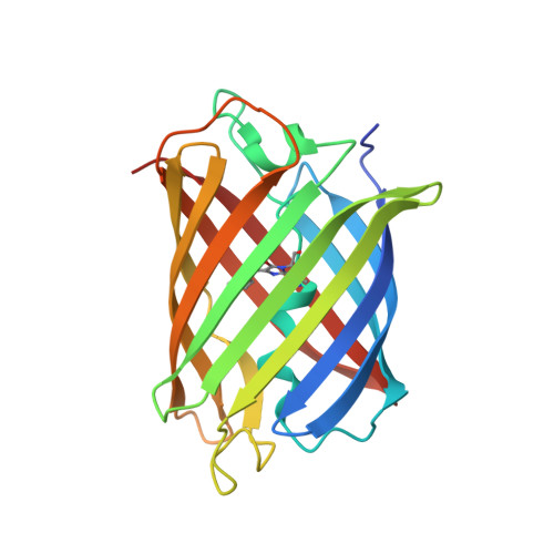 | |
| Modified Residues 1 Unique | |||||
|---|---|---|---|---|---|
| ID | Chains | Type | Formula | 2D Diagram | Parent |
| C12 Query on C12 | A | L-PEPTIDE LINKING | C15 H18 N3 O5 |  | THR, TYR, GLY |
| Length ( Å ) | Angle ( ˚ ) |
|---|---|
| a = 81.156 | α = 90 |
| b = 81.156 | β = 90 |
| c = 84.977 | γ = 90 |
| Software Name | Purpose |
|---|---|
| HKL-2000 | data reduction |
| SCALEPACK | data scaling |
| PHENIX | refinement |
| PDB_EXTRACT | data extraction |
| PHENIX | phasing |
| Funding Organization | Location | Grant Number |
|---|---|---|
| National Institutes of Health/National Institute of General Medical Sciences (NIH/NIGMS) | United States | -- |


