Redox properties and PAS domain structure of the Escherichia coli energy sensor Aer indicate a multistate sensing mechanism.
Maschmann, Z.A., Chua, T.K., Chandrasekaran, S., Ibanez, H., Crane, B.R.(2022) J Biol Chem 298: 102598-102598
- PubMed: 36252616 Search on PubMedSearch on PubMed Central
- DOI: https://doi.org/10.1016/j.jbc.2022.102598
- Primary Citation Related Structures:
8DIK - PubMed Abstract:
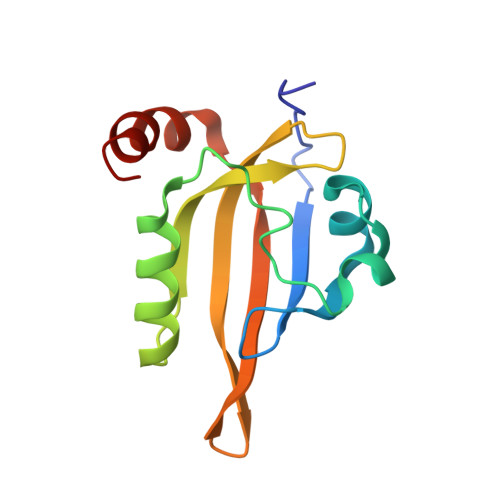
The Per-Arnt-Sim (PAS; named for the representative proteins: Period, Aryl hydrocarbon receptor nuclear translocator protein and Single-minded) domain of the dimeric Escherichia coli aerotaxis receptor Aer monitors cellular respiration through a redox-sensitive flavin adenine dinucleotide (FAD) cofactor. Conformational shifts in the PAS domain instigated by the oxidized FAD (FAD OX )/FAD anionic semiquinone (FAD ASQ ) redox couple traverse the HAMP (histidine kinases, adenylate cyclases, methyl-accepting chemotaxis proteins, and phosphatases) and kinase control domains of the Aer dimer to regulate CheA kinase activity. The PAS domain of Aer is unstable and has not been previously purified. Here, residue substitutions that rescue FAD binding in an FAD binding-deficient full-length Aer variant were used in combination to stabilize the Aer PAS domain. We solved the 2.4 Å resolution crystal structure of this variant, Aer-PAS-GVV, and revealed a PAS fold that contains distinct features associated with FAD-based redox sensing, such as a close contact between the Arg115 side chain and N5 of the isoalloxazine ring and interactions of the flavin with the side chains of His53 and Asn85 that are poised to convey conformational signals from the cofactor to the protein surface. In addition, we determined the FAD ox /FAD ASQ formal potentials of Aer-PAS-GVV and full-length Aer reconstituted into nanodiscs. The Aer redox couple is remarkably low at -289.6 ± 0.4 mV. In conclusion, we propose a model for Aer energy sensing based on the low potential of Aer-PAS-FAD ox /FAD ASQ couple and the inability of Aer-PAS to bind to the fully reduced FAD hydroquinone.
- Department of Chemistry and Chemical Biology, Cornell University, Ithaca, New York, USA.
Organizational Affiliation: